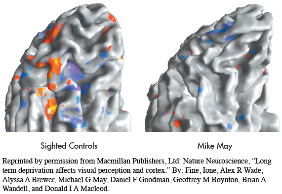
Scanning Mike’s Brain The red, orange, and yellow colors in the left fMRI scan show the areas of the occipital lobe that are normally activated in response to faces. Blue and purple indicate the typical pattern of brain activity in response to objects. In contrast to a normally sighted individual, Mike’s fMRI scan (right), taken three years after his surgery, shows virtually no response to faces and only slight brain activation in response to objects.
Reprinted by permission from Macmillan Publishers, Ltd: Nature Neuroscience, “Long term deprivation affects visual perception and cortex.” By: Fine, Ione, Alex R Wade, Alyssa A Brewer, Michael G May, Daniel F Goodman, Geoffrey M Boynton, Brian A Wandell, and Donald I A Macleod.