4.1 The Tools of Discovery: Having Our Head Examined
4-1 How do neuroscientists study the brain’s connections to behavior and mind?
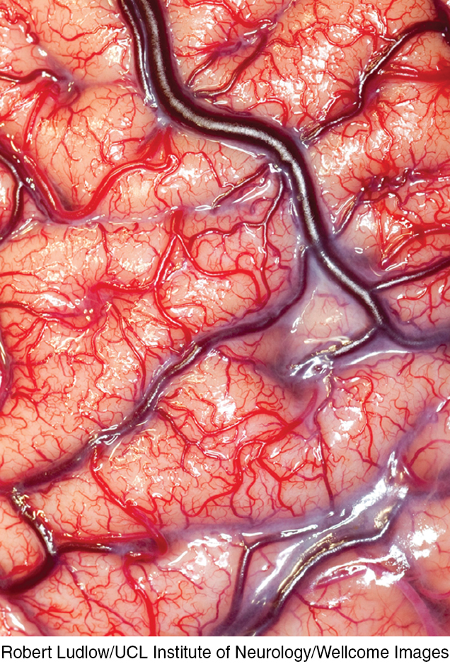
A living human brain exposed Today’s neuroscience tools enable us to “look under the hood” and glimpse the brain at work, enabling the mind.
Robert Ludlow/UCL Institute of Neurology/Wellcome Images
A century ago, scientists had no tools high powered yet gentle enough to explore the living human brain. Early case studies helped localize some brain functions. Damage to one side of the brain often caused numbness or paralysis on the opposite side, suggesting that the body’s right side is wired to the brain’s left side, and vice versa. Damage to the back of the brain disrupted vision, and damage to the left-front part of the brain produced speech difficulties. Gradually, these early explorers were mapping the brain.
lesion [LEE-zhuhn] tissue destruction. A brain lesion is a naturally or experimentally caused destruction of brain tissue.
Now, within a lifetime, a new generation of neural mapmakers is charting the known universe’s most amazing organ. Whether in the interests of science or medicine, they can selectively lesion (destroy) tiny clusters of normal or defective brain cells, leaving the surrounding tissue unharmed. Today’s scientists can snoop on the messages of individual neurons, using modern microelectrodes with tips small enough to detect the electrical pulse in a single neuron. For example, they can now detect exactly where the information goes in a cat’s brain when someone strokes its whisker. They can also stimulate various brain parts and note the effect, eavesdrop on the chatter of billions of neurons, and see color representations of the brain’s energy-consuming activity. These techniques for peering into the thinking, feeling brain are doing for psychology what the microscope did for biology and the telescope did for astronomy.
electroencephalogram (EEG) an amplified recording of the waves of electrical activity sweeping across the brain’s surface. These waves are measured by electrodes placed on the scalp.
Right now, your mental activity is emitting telltale electrical, metabolic, and magnetic signals that would enable neuroscientists to observe your brain at work. Electrical activity in your brain’s billions of neurons sweeps in regular waves across its surface. An electroencephalogram (EEG) is an amplified readout of such waves (FIGURE 4.1). Researchers record the brain waves through a shower-cap-like hat that is filled with electrodes covered with a conductive gel.
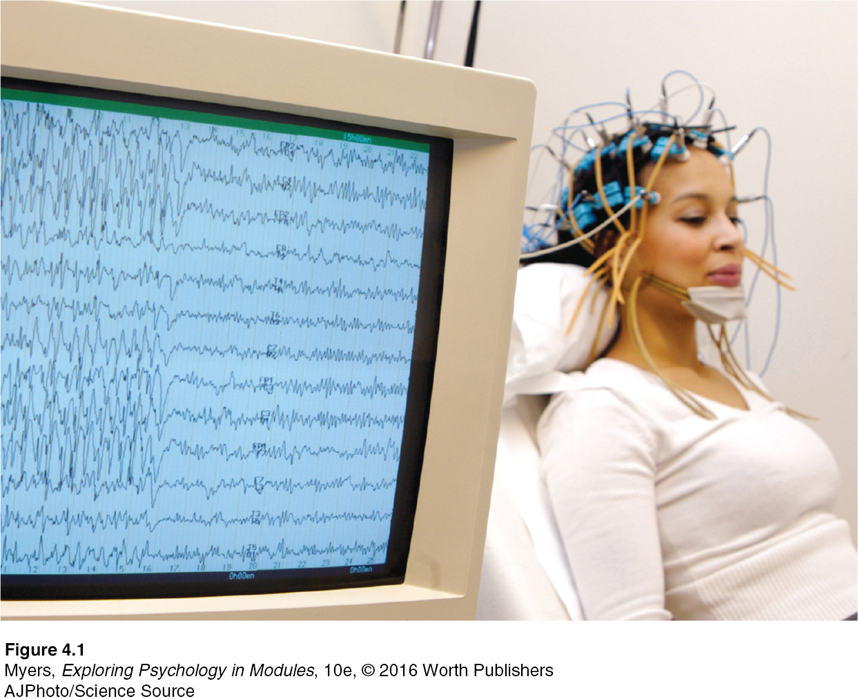
Figure 2.9: FIGURE 4.1 Brain hacking An electroencephalograph provides amplified tracings of waves of electrical activity in the brain.
AJPhoto/Science Source
PET (positron emission tomography) scan a visual display of brain activity that detects where a radioactive form of glucose goes while the brain performs a given task.
“You must look into people, as well as at them,” advised Lord Chesterfield in a 1746 letter to his son. Unlike EEGs, newer neuroimaging techniques give us that Superman-like ability to see inside the living brain. One such tool, the PET (positron emission tomography) scan (FIGURE 4.2), depicts brain activity by showing each brain area’s consumption of its chemical fuel, the sugar glucose. Active neurons are glucose hogs. Our brain, though only about 2 percent of our body weight, consumes 20 percent of our calorie intake. After a person receives temporarily radioactive glucose, the PET scan can track the gamma rays released by this “food for thought” as a task is performed. Rather like weather radar showing rain activity, PET-scan “hot spots” show the most active brain areas as the person does mathematical calculations, looks at images of faces, or daydreams.
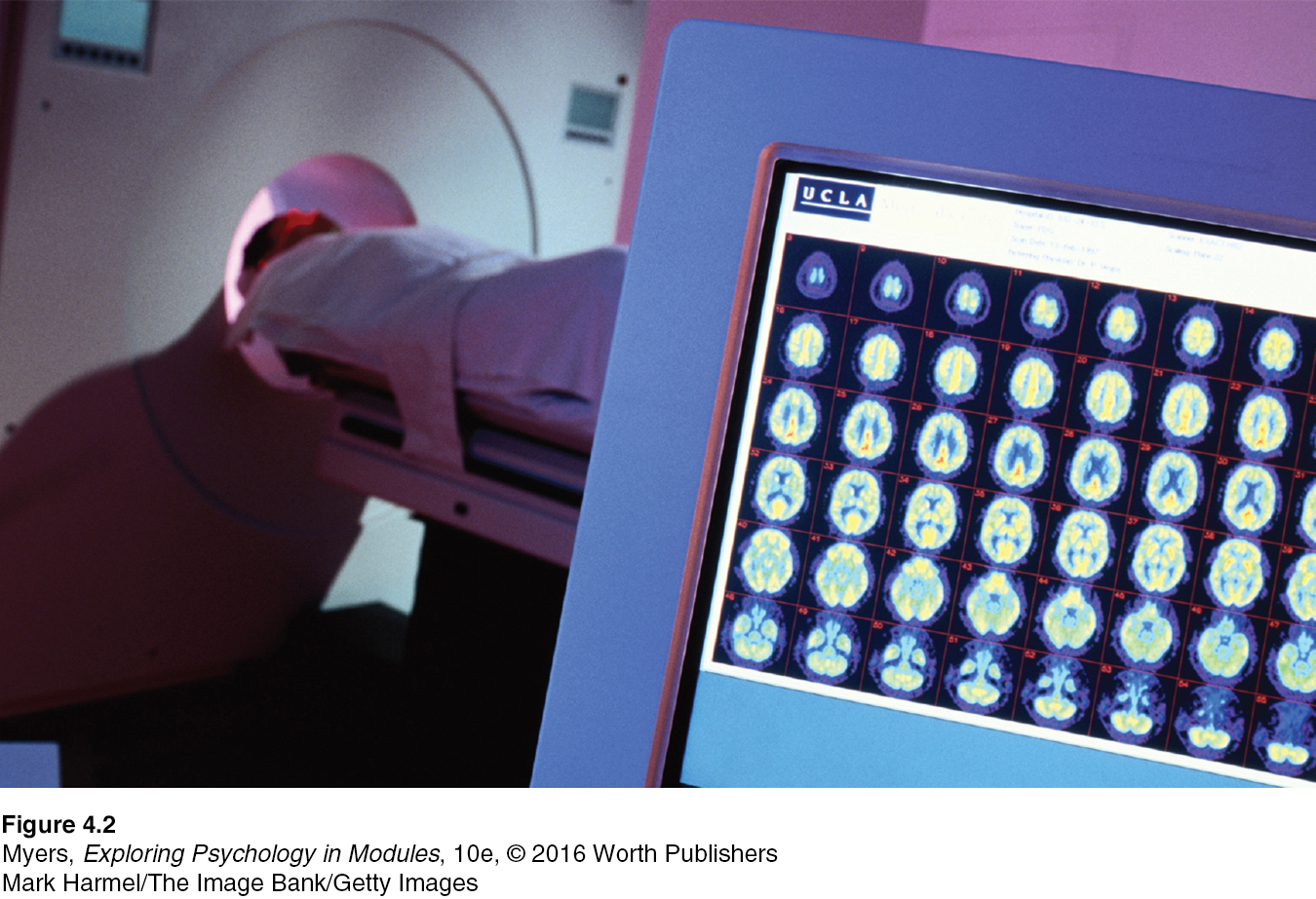
Figure 2.10: FIGURE 4.2 The PET scan To obtain a PET scan, researchers inject volunteers with a low and harmless dose of a short-lived radioactive sugar. Detectors around the person’s head pick up the release of gamma rays from the sugar, which has concentrated in active brain areas. A computer then processes and translates these signals into a MAP of the brain at work.
Mark Harmel/The Image Bank/Getty Images
MRI (magnetic resonance imaging) a technique that uses magnetic fields and radio waves to produce computer-generated images of soft tissue. MRI scans show brain anatomy.
In MRI (magnetic resonance imaging) brain scans, the person’s head is put in a strong magnetic field, which aligns the spinning atoms of brain molecules. Then, a radio-wave pulse momentarily disorients the atoms. When the atoms return to their normal spin, they emit signals that provide a detailed picture of soft tissues, including the brain. MRI scans have revealed a larger-than-average neural area in the left hemisphere of musicians who display perfect pitch (Schlaug et al., 1995). They have also revealed enlarged ventricles—fluid-filled brain areas (marked by the red arrows in FIGURE 4.3)—in some patients who have schizophrenia, a disabling psychological disorder.
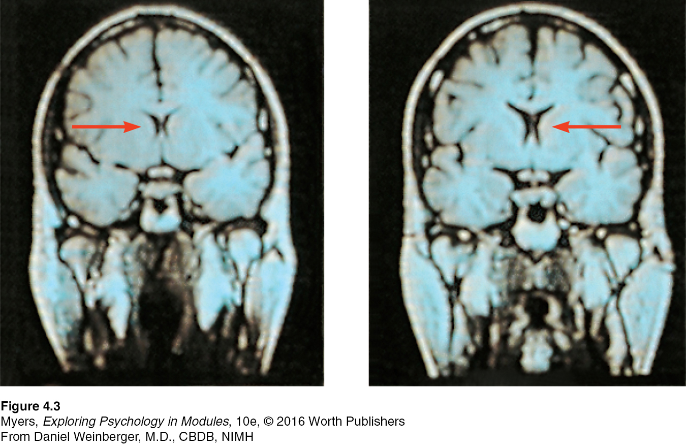
Figure 2.11: FIGURE 4.3 MRI scan of a healthy individual (left) and a person with schizophrenia (right) Note the enlarged ventricle, the fluid-filled brain region at the tip of the arrow in the image on the right.
From Daniel Weinberger, M.D., CBDB, NIMH
fMRI (functional MRI) a technique for revealing bloodflow and, therefore, brain activity by comparing successive MRI scans. fMRI scans show brain function as well as structure.
A special application of MRI—fMRI (functional MRI)—can reveal the brain’s functioning as well as its structure. Where the brain is especially active, blood goes. By comparing successive MRI scans, researchers can watch as specific brain areas activate, showing increased oxygen-laden bloodflow. As a person looks at a scene, for example, the fMRI machine detects blood rushing to the back of the brain, which processes visual information (see FIGURE 5.3). When the brain is unoccupied, blood continues to flow via a web of brain regions called the default network (Mason et al., 2007).
Such snapshots of the brain’s activity provide new insights into how the brain divides its labor. A mountain of recent fMRI studies suggests which brain areas are most active when people feel pain or rejection, listen to angry voices, think about scary things, feel happy, or become sexually excited. The technology enables a very crude sort of mind reading. One neuroscience team scanned 129 people’s brains as they did eight different mental tasks (such as reading, gambling, or rhyming). Later, they were able, with 80 percent accuracy, to predict which of these mental activities their participants had been doing (Poldrack et al., 2009).
You’ve seen the pictures—of colorful brains with accompanying headlines, such as “your brain on music.” Hot brains make hot news (Fine, 2010). But “neuroskeptics” caution against overblown claims (Satel & Lilienfeld, 2013; Vul et al., 2009a,b). Neuromarketing, neuropolitics, and neurotheology are often neurohype. Imaging techniques illuminate brain structure and activity, and sometimes help us test different theories of behavior (Mather et al., 2013). But given that all human experience is brain-based, it’s no surprise that different brain areas become active when one listens to a lecture or lusts for a lover.
Nevertheless, to learn about the neurosciences now is like studying world geography when Magellan explored the seas. The $40 million Human Connectome Project (2013; Gorman, 2014), for example, seeks “neural pathways [that] will reveal much about what makes us uniquely human and what makes every person different from all others.” It harnesses the power of diffusion spectrum imaging, a type of MRI technology that maps long-distance brain fiber connections (Jarbo & Verstynen, 2015). Today’s whole-brain mapping effort has been likened to last century’s Apollo program, which landed humans on the Moon. This truly is the golden age of brain science.
RETRIEVE IT
Match the scanning technique with the correct description.
Question
q/Egz9HHVa0Um9Zalc++JpO1TttPLRGUn1tdsYP5u9u7fc1gJGOBkAHzwY32l09pCkaeNhO9l5c6zpv1eLCxSRc27/T3YnYjOPrzIkXcx49i+4/w9sPezUIgVxtGXxna90ZkLRqaiX9kRwog6cJOsK0CE9B+PmvsbGteCqW9aqrghmz7v7u6/qzLJzIrBi5gdKKWtX5/tjS2J6KHpsAhdgArV3Mih1rGXMe/75bF6kfSuaiTK6voHkMqulNlHA/0CMcC2I7Mh+ZblWNIqvgRrNDqaRGXLwEVemwNMQ==