4.2 Older Brain Structures
4-
 For an introductory 12.5-
An animal’s capacities come from its brain structures. In primitive animals, such as sharks, a not-
This increasing complexity arises from new brain systems built on top of the old, much as Earth’s landscape covers the old with the new. Digging down, one discovers the fossil remnants of the past—
The Brainstem
brainstem the oldest part and central core of the brain, beginning where the spinal cord swells as it enters the skull; the brainstem is responsible for automatic survival functions.
medulla [muh-
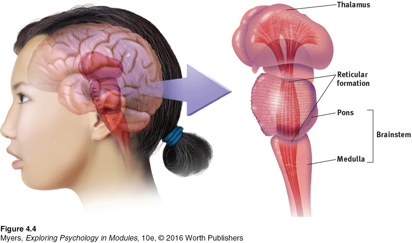
The brain’s oldest and innermost region is the brainstem. It begins where the spinal cord swells slightly after entering the skull. This slight swelling is the medulla (FIGURE 4.4). Here lie the controls for your heartbeat and breathing. As brain-
51
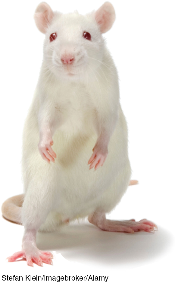
If a cat’s brainstem is severed from the rest of the brain above it, the animal will still breathe and live—
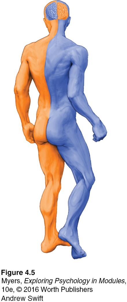
The brainstem is a crossover point, where most nerves to and from each side of the brain connect with the body’s opposite side (FIGURE 4.5). This peculiar cross-
RETRIEVE IT
Question
Nerves from the left side of the brain are mostly linked to the le2nixNaZOa02/4j side of the body, and vice versa.
The Thalamus
thalamus [THAL-
Sitting atop the brainstem is the thalamus, a pair of egg-
The Reticular Formation
52
reticular formation a nerve network that travels through the brainstem into the thalamus and plays an important role in controlling arousal.
Inside the brainstem, between your ears, lies the reticular (“netlike”) formation, a neuron network extending from the spinal cord right up through the thalamus. As the spinal cord’s sensory input flows up to the thalamus, some of it travels through the reticular formation, which filters incoming stimuli, relays important information to other brain areas, and controls arousal.
In 1949, Giuseppe Moruzzi and Horace Magoun discovered that electrically stimulating a sleeping cat’s reticular formation almost instantly produced an awake, alert animal. When Magoun severed a cat’s reticular formation without damaging nearby sensory pathways, the effect was equally dramatic: The cat lapsed into a coma from which it never awakened.
The Cerebellum
cerebellum [sehr-
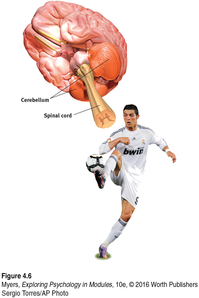
Extending from the rear of the brainstem is the baseball-
* * *
Note: These older brain functions all occur without any conscious effort. This illustrates another of our recurring themes: Our brain processes most information outside of our awareness. We are aware of the results of our brain’s labor—
RETRIEVE IT
Question
T6VXbJ3hstFF4fY07YvrZiuPf+RcVTZbLS7oJoBP4Y9CgSIRNTAuQ+/mq6WtrlytrmOT66HPec6Irn40InJWxAwqpD08O3mTSbmQ1E61depfYUQXLBCoU5Lu4i+YyfzXyIiW4R67MJbzJ40QbDrgTMpiGPcGGu2b5AOBwVJ0sRrTxhcl2V47z8fXo8P5B+mVfltayKXkki4yRa2HAi1KXLTJ6SPdYylajK1hMEcZoHdPUXrlOdJYekbHjtkosnJ2NEbH5inbu6HStf8oODiZZFMKI8mEDiay67dHgYQy+GE=The Limbic System
limbic system neural system (including the amygdala, hypothalamus, and hippocampus) located below the cerebral hemispheres; associated with emotions and drives.
4-
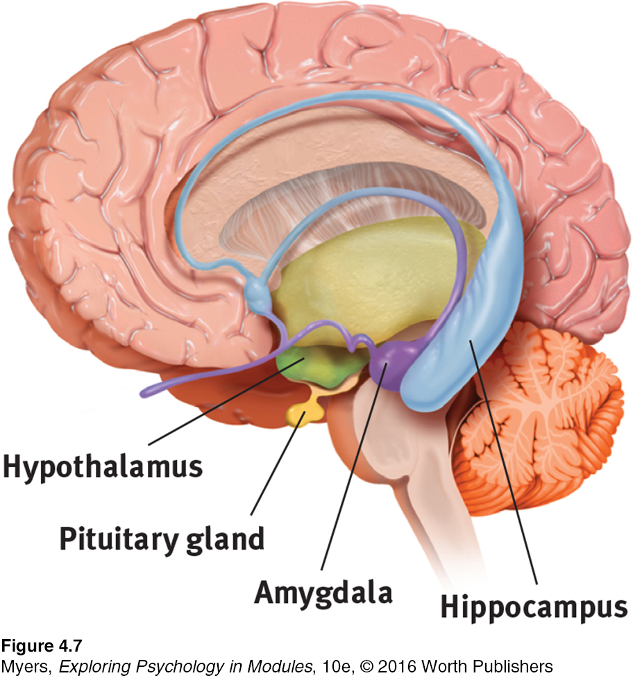
We’ve considered the brain’s oldest parts, but we’ve not yet reached its newest and highest regions, the cerebral hemispheres (the two halves of the brain). Between the oldest and newest brain areas lies the limbic system (limbus means “border”). This system contains the amygdala, the hypothalamus, and the hippocampus (FIGURE 4.7).
amygdala [uh-
THE AMYGDALA Research has linked the amygdala, two lima-
53
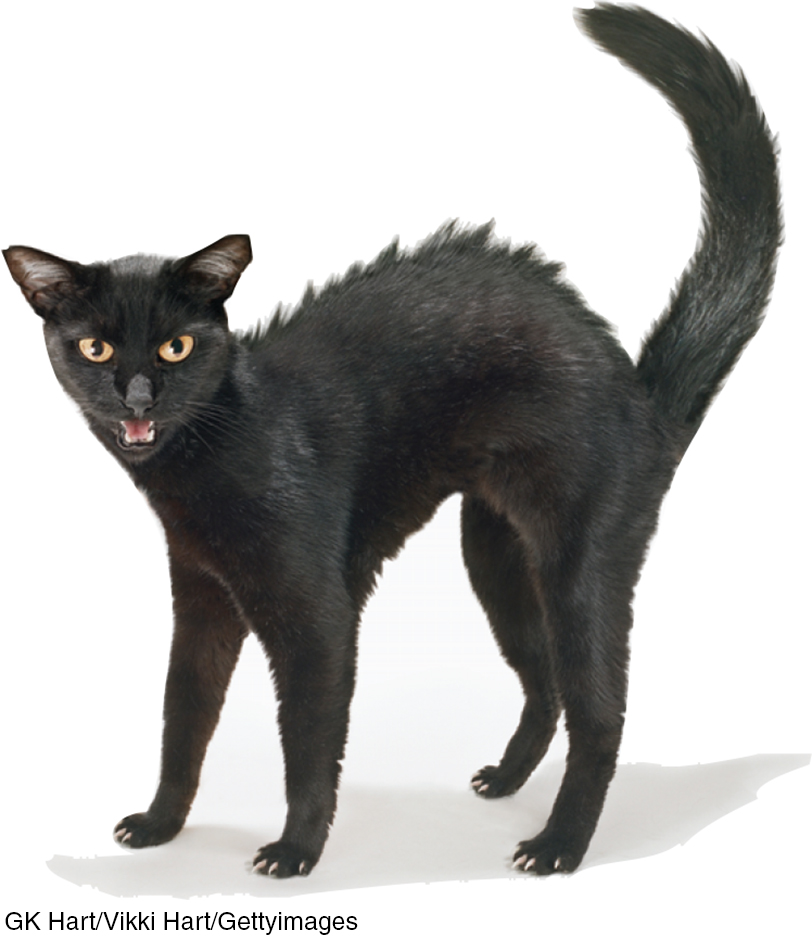
What then might happen if we electrically stimulated the amygdala of a normally placid domestic animal, such as a cat? Do so in one spot and the cat prepares to attack, hissing with its back arched, its pupils dilated, its hair on end. Move the electrode only slightly within the amygdala, cage the cat with a small mouse, and now it cowers in terror.
These and other experiments have confirmed the amygdala’s role in fear and rage. One study found math anxiety associated with hyperactivity in the right amygdala (Young et al., 2012). Other studies link criminal behavior with amygdala dysfunction (Boccardi et al., 2011; Ermer et al., 2012). But we must be careful. The brain is not neatly organized into structures that correspond to our behavior categories. When we feel or act in aggressive or fearful ways, there is neural activity in many areas of our brain—
RETRIEVE IT
Question
2ZjrqsOtVCxfeeOCkYFY2/emvNI+hFf/PxyX6WwMFdkCkzde+oFS2OwZveaE2H0BaKa8TkHgHyIcxt08EyhmvHU+oNP64DfAHycomQG70ITnc/hJkiz/7IVTv2h16SyENptX90PtFmpxwr2Sat94pzx8mp3H0IkxnvsgVS5iDtq9Ju3ZEcnfdPysYmyqBOFSsqwN6G5zz23DrFkPhypothalamus [hi-
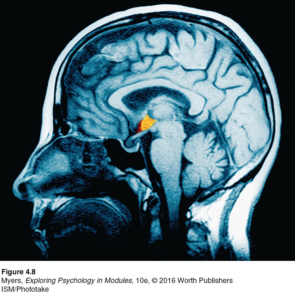
THE HYPOTHALAMUS Just below (hypo) the thalamus is the hypothalamus (FIGURE 4.8), an important link in the command chain governing bodily maintenance. Some neural clusters in the hypothalamus influence hunger; others regulate thirst, body temperature, and sexual behavior. Together, they help maintain a steady (homeostatic) internal state.
As the hypothalamus monitors the state of your body, it tunes into your blood chemistry and any incoming orders from other brain parts. For example, picking up signals from your brain’s cerebral cortex that you are thinking about sex, your hypothalamus will secrete hormones. These hormones will in turn trigger the adjacent “master gland” of the endocrine system, your pituitary (see FIGURE 4.7), to influence your sex glands to release their hormones. These will intensify the thoughts of sex in your cerebral cortex. (Once again, we see the interplay between the nervous and endocrine systems: The brain influences the endocrine system, which in turn influences the brain.)
A remarkable discovery about the hypothalamus illustrates how progress in science often occurs—
Later experiments located other “pleasure centers” (Olds, 1958). (What the rats actually experience only they know, and they aren’t telling. Rather than attribute human feelings to rats, today’s scientists refer to reward centers, not “pleasure centers.”) Just how rewarding are these reward centers? Enough to cause rats to self-
54
Do humans have limbic centers for pleasure? To calm violent patients, one neurosurgeon implanted electrodes in such areas. Stimulated patients reported mild pleasure; unlike Olds’ rats, however, they were not driven to a frenzy (Deutsch, 1972; Hooper & Teresi, 1986). Moreover, newer research reveals that stimulating the brain’s “hedonic hotspots” (its reward circuits) produces more desire than pure enjoyment (Kringelbach & Berridge, 2012).
“If you were designing a robot vehicle to walk into the future and survive, . . . you’d wire it up so that behavior that ensured the survival of the self or the species—
Candace Pert (1986)
Some researchers believe that substance use disorders may stem from malfunctions in natural brain systems for pleasure and well-
hippocampus a neural center located in the limbic system; helps process explicit memories for storage.
THE HIPPOCAMPUS The hippocampus processes conscious, explicit memories and decreases in size and function as we grow older. Animals or humans who lose their hippocampus to surgery or injury lose their ability to form new memories of facts and events. Those who survive a hippocampal brain tumor in childhood struggle to remember new information in adulthood (Jayakar et al., 2015). Other modules discuss how hippocampus size and function decrease as we grow older and how our two-
* * *
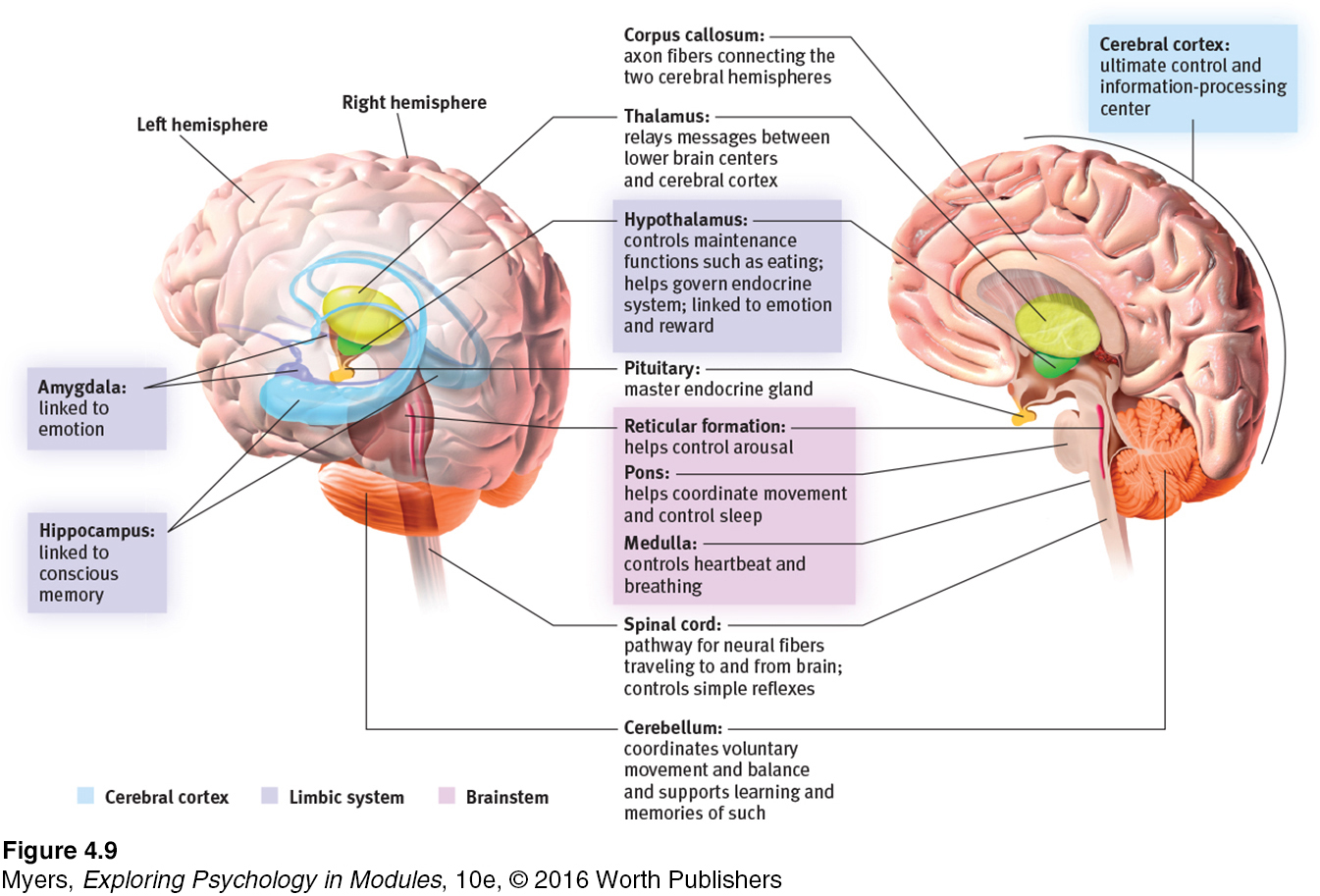
FIGURE 4.9 locates the brain areas we’ve discussed, as well as the cerebral cortex, our next topic.
55
To review and assess your understanding, visit LaunchPad’s Concept Practice: The Limbic System.