5.2 Methods of Mapping the Brain’s Behavioral Functions
With its billions of neurons and trillions of synapses, the brain is a daunting subject of study. It would be hopeless, with present technology, to try to work out all of the details of its wiring, as one might with a human-made machine such as a radio or a computer. Fortunately, though, for those of us who want to understand as much as we can of it, patterns exist in the brain’s trillions of connections.
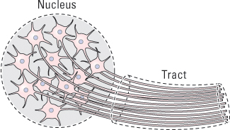
Neurons in the central nervous system are organized into nuclei and tracts. A nucleus is a cluster of cell bodies in the central nervous system (not to be confused with the cell nucleus, which exists within each cell), and a tract is a bundle of axons that course together from one nucleus to another (see Figure 5.11). A tract in the central nervous system is analogous to a nerve in the peripheral nervous system. Tracts are referred to collectively as white matter because the myelin sheaths around axons make tracts appear relatively white; nuclei, which are darker, are referred to collectively as gray matter.
In general, neurons whose cell bodies occupy the same nucleus and whose axons run in the same tract have identical or similar functions, and groups of nuclei located near one another have functions that are closely related to one another. Because of this pattern of organization, we can speak of the general functions of relatively large anatomical structures within the brain.
Methods Used for Studying the Human Brain
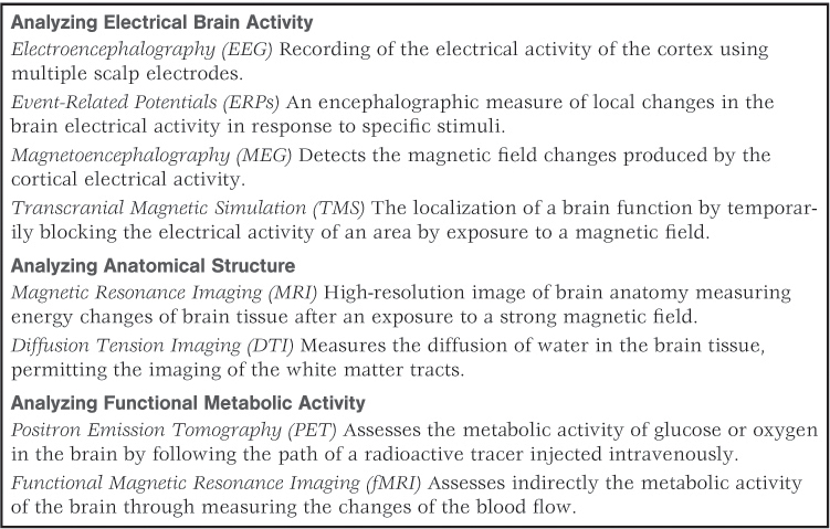
Psychologists and other neuroscientists have developed a number of methods of identifying the functions of specific brain areas. The methods fall into three general categories: (1) observing behavioral deficits that occur when a part of the brain is destroyed or is temporarily inactivated; (2) observing behavioral effects of artificially stimulating specific parts of the brain; and (3) recording changes in neural activity that occur in specific parts of the brain when a person or animal is engaged in a particular mental or behavioral task. Table 5.1 provides a partial list of some of the imaging techniques in use today. What follows are brief descriptions of the most common methods used within each of these three categories. We’ll look first at methods used in studying the brains of humans and then, in the next section, at more intrusive methods used in studying the brains of nonhuman animals.
Some techniques for studying the brain
160
Observing Effects of Localized Brain Damage
10
How do researchers identify functions of areas of the human brain through (a) studying the effects of brain damage, (b) using a magnetic field to interrupt normal brain activity, (c) recording electrical activity that passes through the skull and scalp, and (d) creating images that depict patterns of blood flow?
At the height of his career, the Russian composer V. G. Shebalin suffered a stroke (a rupturing of blood vessels in the brain) that damaged a portion of his left cerebral cortex (the highest outermost part of the brain). From then on, he had great difficulty expressing himself in words or understanding what others were saying, but he continued to create great music. His Fifth Symphony, composed after his stroke, was described by the composer Dmitri Shostakovich as “a brilliant work, filled with highest emotions, optimistic and full of life” (Gardner, 1982, p. 327).
As illustrated by this case, brain damage often leaves a person deficient in some areas of mental functioning yet fully capable, or nearly so, in others. By studying many people who have damage in the same general area of the brain, psychologists can draw reasonable inferences about the behavioral and mental functions of that area. The logic here is that if some abilities are missing and other abilities remain when a part of the brain is missing, then that brain part must contribute in some essential way to the missing abilities but not to those that remain. As you will discover later in this chapter and elsewhere in the book, much has been learned about the human brain through systematic studies of people who have suffered brain damage.
However, one must be cautious when interpreting the effects of brain damage on psychological functioning. Brain damage can rarely be narrowed to one area and frequently involves complications beyond that of simple lesions, or areas of specific damage. Lesions in one area of the brain can also lead to changes in other brain areas (Fuster, 1989). Despite these and other reservations, much can be learned about brain functioning from studying brain damage, especially when viewed in combination with other sources of data.
Observing Effects of Magnetic Interference with Normal Brain Activity
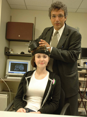
A relatively new procedure for localizing functions in the human brain is transcranial magnetic stimulation, or TMS (Pascual-Leone et al., 2005). In this procedure, a pulse of electricity is sent through a small copper coil, inducing a magnetic field around the coil. The coil is held just above a person’s scalp so that the magnetic field passes through the scalp and skull and induces an electric current in the neurons immediately below the coil. Repetitive pulses cause a temporary loss in those neurons’ abilities to fire normally. The effect is comparable to that of lesioning a small area of the brain, with the advantage that the effect is temporary and reversible. For instance, if the coil is held over an area of the brain that is crucial for fluent speech, the person becomes unable to speak fluently while the current is on, but then resumes fluent speech soon after the current is turned off. Because the magnetic field affects only that part of the brain lying immediately below the skull, TMS can be used for mapping the functions only of the outermost yet largest part of the brain, the cerebral cortex.
A good example of research using TMS is found in the work of Sara Torriero and her colleagues (2007) in Italy. These researchers were interested in the neural foundations of observational learning (learning from watching others). In one of these experiments, subjects observed another person solve a problem in which a series of squares had to be pressed in a specified order. Different groups of subjects received TMS over different parts of the brain just prior to the observation. The result was that those who received the TMS directly over a portion of the brain called the dorsolateral prefrontal cortex failed to benefit from observing the problem’s solution. Even though the TMS did not interfere with their vision, it took these subjects as long to learn the sequence they had observed—by trial and error—as it took them to learn a new sequence they had never observed. This study now constitutes part of the evidence that a system of mirror neurons (discussed earlier in this chapter) exists in this part of the cerebral cortex, which is crucial for translating observed actions into self-produced actions.
161
Although TMS is usually used to study the effects of temporary inactivation of a brain area, it can also be used to study the effects of temporary activation. A single pulse of electrical current can cause the sets of neurons just below the coil to send a burst of action potentials through their axons. When the coil is held over an area of the brain responsible for controlling the body’s muscle movements, the result is a brief muscle twitch somewhere in the body—such as in the thumb of one hand. In this way, TMS can be used to produce a map showing the functional connections between specific areas within movement-control portions of the cerebral cortex and the muscles controlled by those areas.
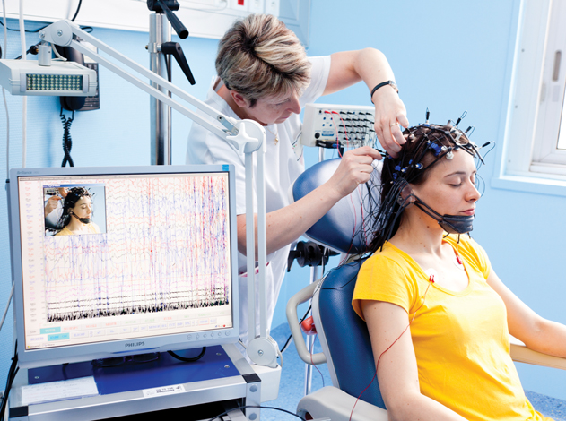
Recording Brain Activity Electrically, Through the Scalp
The constant activity of the brain’s billions of neurons produces continuous electrical “chatter,” which to some degree penetrates the overlying skull and scalp. By placing electrodes on a person’s scalp, researchers can detect and amplify these signals. The resulting record of brain activity is referred to as an electroencephalogram [i-lěk´-trō-in-sěf´-ǝ-lō-grăm] (abbreviated EEG). Patterns in the EEG can be used as an index of whether a person is highly aroused, or relaxed, or asleep and can be used to identify various stages of sleep.
With computer technology, it is possible to record and analyze brain activity simultaneously from many electrodes, each at a different location on the scalp, to see how brain activity varies from place to place as well as from instant to instant (see Figure 5.12). In some of these studies, subjects are asked to respond to stimuli that are presented to them as their brain activity is being recorded. For instance, a person might be presented with sets of letters flashed on a screen and instructed to push a button if and only if the letters spell out an English word. The brief change in the EEG record immediately following the stimulus is referred to as an event-related potential, or ERP. By testing the person repeatedly in such a task and averaging the EEG records obtained, it is possible to produce an average ERP for each electrode location. Comparison of the average ERPs recorded at different scalp locations reveals the pattern of activity in the brain as the person detects and responds to the stimulus. For the task just described, areas of the brain that are involved in seeing the letters would respond first, and areas that are involved in reading and making judgments would respond a split second later.
Viewing Brain Activity with Imaging Methods Sensitive to Blood Flow
Some methods for localizing brain activity rely on the fact that increased neural activity in any area of the brain is accompanied by increased blood flow to that area. Like other bodily tissues, the brain is permeated by tiny blood vessels. When a portion of the brain becomes more active, blood vessels there immediately enlarge, so more blood enters that portion. The blood carries oxygen and glucose, which are the sources of energy required to sustain the increased neural activity. Using technically complex methods, researchers can create three-dimensional pictures, referred to as neuroimages, that depict the relative amount of blood flowing through each part of the brain. The assumption, now well validated, is that increased blood flow reflects increased neural activity (Logothetis, 2008).
162
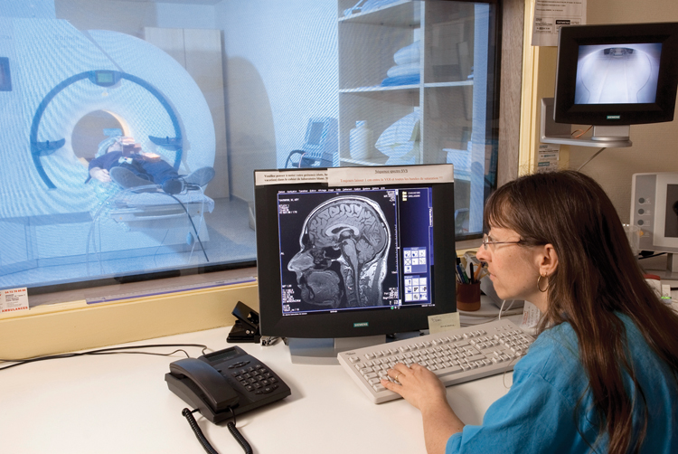
The first of these neuroimaging methods (developed in the 1970s) was positron emission tomography [tō-mȯg´-rǝ-fē], or PET. This method involves injecting a radioactive substance into the blood (in an amount that is not dangerous to the subject) and measuring the radioactivity that is emitted from each portion of the brain. Another method (first used extensively in the 1990s) is functional magnetic resonance imaging, or fMRI. This method involves the creation of a magnetic field around a person’s head, which causes hemoglobin molecules that are carrying oxygen in the blood to give off radio waves of a certain frequency, which can be detected and used to assess the amount of blood in each part of the brain. With either method, the person’s head must be surrounded by a set of sensors and, with fMRI, also by magnetic coils (see Figure 5.13). During the procedure, the person can communicate with the experimenter through a microphone and can respond to visual stimuli presented on a screen inside the scanning device. With either PET or fMRI, a computer is used to generate a three-dimensional image of the brain that depicts variations in amount of blood flow as variations in color (see Figure 5.14).
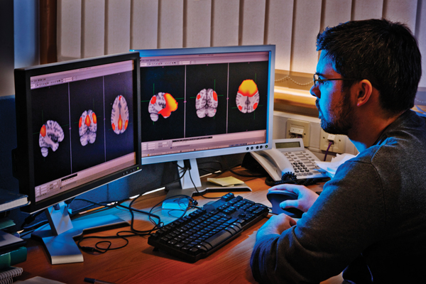
Unlike the EEG, both PET and fMRI can depict activity anywhere in the brain, not just on the surface near the skull. These methods also produce a more fine-grained picture of the spatial locations of activity than is possible with EEG. Today fMRI is used much more often than PET, partly because it shows better spatial resolution.
At any given time, the brain is doing many things, and all portions of it are always active to some degree. (Don’t believe the often-heard statement that people only use 10% of their brains! People use all of their brains all of the time.) In using either PET or fMRI to determine which brain areas are most directly involved in a particular task, researchers need to employ an appropriate control condition. By subtracting the amount of activity measured in each brain area in the control condition (when the person is not performing the specific task) from the amount measured in the same areas in the experimental condition (when the person is performing the task), the researcher can determine which areas show the greatest increase in activity during the task. (For an example of such an experiment, and to see sample PET images, look ahead to Figure 5.33 on p. 185.)
There are, of course, limitations to such methods, and we must not confuse knowing what areas “light up” when a subject performs a particular task (recalling a word, for example) with actually understanding what the neurological and psychological processes are that underlie the action. For example, if we know that the brain areas that “light up” in fMRIs of children with intellectual disabilities are different from those that light up in fMRIs of typically developing children, this by itself does not tell us anything about the origins of the impairment or how to deal with such children. Yet there is a tendency for the public, including policy makers, to believe that the results of psychological studies involving brain images are more convincing than studies without brain images (Beck, 2010). The new technologies of neuroimaging are providing a window into the brain that can help us get a better understanding of human thought and behavior, but such results will be most useful when accompanied by behavioral assessment and experimental studies (Decety & Cacioppo, 2010).
163
Methods Used for Studying the Brains of Nonhuman Animals
With nonhuman animals, researchers can localize brain functions using methods that are more intrusive than those used with humans. They can destroy, stimulate, or record neural activity in small, well-localized areas anywhere in the brain in order to assess the behavioral functions of those areas.
Observing Effects of Deliberately Placed Brain Lesions
11
How do researchers damage, stimulate, and record from neurons in specific areas of animal brains to learn about the functions of those brain areas?
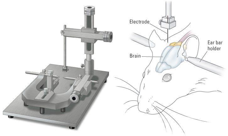
Areas of damage (referred to as lesions) can be produced in the brains of rats or other laboratory animals by either electrical or chemical means. To produce a lesion electrically, a thin wire electrode, insulated everywhere except at its tip, is surgically inserted into the brain with the help of a stereotaxic instrument (see Figure 5.15), and enough current is sent through the electrode to destroy the neurons adjacent to its tip. To produce a lesion chemically, a tiny tube called a cannula is inserted into the brain, again using a stereotaxic instrument, and a small amount of a chemical is injected through the cannula, destroying neurons whose cell bodies are located near the cannula’s tip. The lesions produced by such means can be as small as one-fourth of a millimeter in diameter. By varying the precise location of the damage in different groups of animals, and then comparing the groups in behavioral tests, researchers can identify precise areas of the brain that are crucial for specific types of behaviors.
In most such studies, the lesions are made in deep, primitive areas of the brain, areas whose functions are similar for all mammals, including humans. Such studies have helped researchers identify specific brain nuclei that are crucial for basic motivational and emotional states, such as hunger, sexual drive, and fear—topics we will discuss in Chapter 6.
Effects of Stimulating Specific Areas of the Brain
Stimulation of specific brain areas can also be accomplished either electrically or chemically. To stimulate neurons electrically, a wire electrode is lowered into the brain using the same surgical method as that used to produce a lesion. The electrode is then cemented in place (as shown in Figure 5.15) and can be activated at any time after surgery through a wire connection or by radio waves. The electrical current used for stimulation is much weaker than that used for producing a lesion; it is strong enough to induce action potentials in, but not destroy, neurons near the electrode tip. To stimulate neurons chemically, a cannula is permanently implanted in the brain, and shortly before behavioral testing a tiny amount of a neurotransmitter substance or other chemical known to activate neurons is injected through it.
164
Electrical or chemical stimulation in certain deep areas of the brain can cause an animal to exhibit drive states or emotional states that last only as long as the period of stimulation. For instance, stimulation in one area causes a previously sated animal to eat, and stimulation in another area causes a previously calm animal to behave as if frightened, as long as the stimulation continues.
Electrical Recording from Single Neurons
Electrodes can be used not just to destroy or stimulate specific brain areas, but also to record neural activity in specific areas as the animal engages in some behavioral task. Extremely thin microelectrodes, with very fine points that can penetrate into the cell bodies of single neurons, can be permanently implanted in the brain. In some experiments many such electrodes are inserted at once, each able to record the activity of a different single neuron.
Experiments using this technique have revealed some remarkable correlations between an animal’s behavior and the rate of action potentials produced by individual neurons. For example, certain neurons in one area of the brain (the hippocampus, discussed later in the chapter) fire rapidly when, and only when, the animal (usually a rat) faces in a specific direction within the environment in which it is tested (Best et al., 2001; Moser et al., 2008). If the entire test environment (such as an enclosed box with a maze in it) is rotated, the preferred orientations of these neurons rotate too. Thus, a cell that originally fired whenever the animal faced north will fire whenever the animal faces south after the apparatus is turned 180 degrees. Apparently, these neurons, called place cells, help animals keep track of the direction they are facing within familiar environments. If the area of the brain in which these cells are located is destroyed, animals lose their ability to find their way through mazes or to locate objects easily in environments that they have explored.
SECTION REVIEW
Mapping the brain’s behavioral functions.
Structure and Function
- Neurons in a given nucleus and tract have the same or similar functions, and groups of neighboring nuclei generally have closely related functions.
- To study a brain area’s function, we can observe behavioral effects of inactivating or artificially stimulating that area, or we can measure its neural activity during behavioral tasks.
Human Studies
- Studying the effects of existing localized brain damage can aid identification of the functions associated with the damaged area.
- TMS temporarily disrupts or briefly triggers activity in specific cortical areas by means of a magnetic field.
- An EEG records gross electrical activity in areas of the brain just beneath the skull from electrodes placed on the scalp.
- PET and fMRI produce images that depict changes in neural activity in each area of the brain by measuring changes in blood flow.
Animal Studies
- Precisely placed lesions in the brain can be created electrically or chemically to see how damage affects behavior.
- Electrical or chemical stimulation of a specific brain area can help to reveal its functions.
- Microelectrodes permit the recording of electrical activity in single neurons.