5.4 Asymmetry of Higher Functions of the Cerebral Cortex
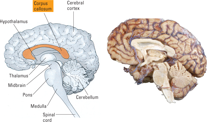
Nearly every part of the brain exists in duplicate. We have a right and a left member of each anatomical portion of the brainstem, thalamus, cerebellum, and so on. The part of the brain in which the right-left division is most evident, however, is the cerebral cortex. Each half of the cortex folds inward where it would abut the other half, forming a deep, fore-to-aft midline fissure that divides the cortex into distinct right and left hemispheres. The two hemispheres are connected, however, by a massive bundle of axons called the corpus callosum [ka-lō-som], which is located below that fissure (see Figure 5.28).
28
In what ways are the two hemispheres of the cerebral cortex symmetrical, and in what ways are they asymmetrical?
The two hemispheres are quite symmetrical in their primary sensory and motor functions. Each does the same job, but for a different half of the body. Most of the neural paths between the primary sensory and motor areas of the cortex and the parts of the body to which they connect are crossed, or contralateral. Thus, sensory neurons that arise from the skin on the right side of the body send their signals to the somatosensory area of the left hemisphere, and vice versa. Similarly, neurons in the primary motor area of the left hemisphere send their signals to muscles on the right side of the body, and vice versa. Such symmetry breaks down, however, in the association areas.
The most obvious distinction between the two cortical hemispheres in humans is that large areas in the left are specialized for language and comparable areas in the right are specialized for nonverbal, visuospatial analysis of information. The earliest evidence of this difference came from observing people who had suffered strokes or other injuries that affected just one hemisphere. In general, damage to the left hemisphere results in deficits in using and comprehending language, and damage to the right results in deficits in such tasks as recognizing faces, reading maps, and drawing geometric shapes, all of which depend on perceiving spatial relationships.
Effects of Surgical Separation of the Hemispheres: Split Brain, Split Mind
Dramatic further evidence of the separate abilities of the two hemispheres appeared in the 1960s, when Roger Sperry, Michael Gazzaniga, and their colleagues began to study patients with “split brains”: people who, as a last-resort treatment for epilepsy, had undergone an operation in which the two hemispheres were separated by cutting the corpus callosum.
181
Earlier, more casual observations had revealed no remarkable deficits in people who had undergone this operation. The operation was generally successful in reducing or eliminating epileptic seizures, and, after a period of recovery, there is usually no drop in measured IQ, in ability to carry on conversations, or even in ability to coordinate the two sides of the body in skilled tasks. But Gazzaniga (1967, 1998) showed that under special test conditions, in which information is provided to just one hemisphere or the other, patients with split brains behave as though they have two separate minds with different abilities.
How Each Hemisphere Can Be Tested Separately After the Split-Brain Operation
29
How is it possible to test each hemisphere separately in people whose corpus callosum has been cut? How do such tests confirm that the left hemisphere controls speech and the right hemisphere has superior spatial ability?
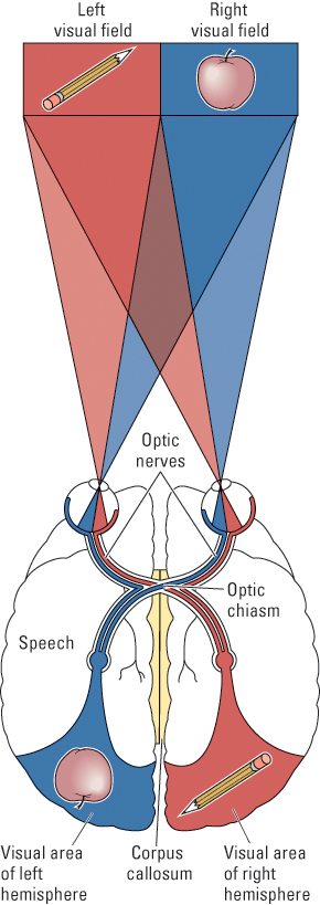
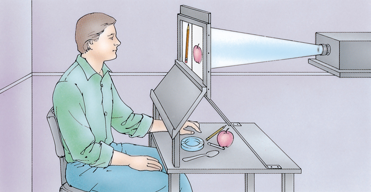
Split-brain studies take advantage of the crossed sensory and motor connections between the brain and peripheral parts of the body. Recall that each hemisphere most directly controls movement in, and receives somatosensory information from, the opposite half of the body. In addition, as illustrated in Figure 5.29, connections from the eyes to the brain are such that input from the right visual field (the right-hand half of a person’s field of view) goes first to the left hemisphere and input from the left visual field goes first to the right hemisphere. In the normal brain, all information that goes to either hemisphere subsequently travels to the other through the corpus callosum. But the split-brain operation destroys those connections. Therefore, with the testing apparatus shown in Figure 5.30 and with patients with split brains as subjects, it is possible to (a) send visual information to just one hemisphere by presenting the stimulus in only the opposite half of the visual field, (b) send tactile (touch) information to just one hemisphere by having the subject feel an object with the opposite hand, and (c) test the knowledge of just one hemisphere by having the subject respond with the hand opposite to that hemisphere.
Split-Brain Evidence for Left-Hemisphere Language and Right-Hemisphere Spatial Ability
In a typical experiment, Gazzaniga flashed pictures of common objects to either the right or the left visual field of a patient with a split brain (see Figure 5.30). When a picture was flashed in the right field (projecting to the left hemisphere), the patient described it as well as someone with an intact brain would; but when a picture was flashed in the left field (projecting to the right hemisphere), the patient either claimed to see nothing or made a random guess. Then Gazzaniga asked the same person to reach under a barrier with one hand or the other and identify, by touch, the object that had been flashed. The fascinating result was that the person could reliably identify with the left hand (but not with the right) the same object that he or she had just vocally denied having seen (Gazzaniga, 1967). For example, if the object flashed to the right hemisphere was a pencil, the subject’s left hand picked out the pencil from a set of objects even while the subject’s voice was continuing to say that nothing had been flashed. Such findings are consistent with the conclusion that only the left hemisphere can generate speech and that neither hemisphere has direct access to what the other one knows.
Research of this sort also revealed large individual differences among patients with split brains in the degree to which the right hemisphere can comprehend speech. Some show essentially no right-hemisphere comprehension. They cannot participate in experiments such as that just described because their right hemispheres don’t understand such instructions as “Pick up the object that you saw on the screen.” Some have reasonably good right-hemisphere comprehension, although their right hemisphere is still unable to generate speech; and a few show a reversal, with the right hemisphere superior to the left in language comprehension and production. Other tests, on people whose corpus callosum has not been cut, have shown that about 4 percent of right-handed individuals and 15 percent of left-handed individuals have their speech centers located in the right hemisphere rather than the left (Rasmussen & Milner, 1977).
182
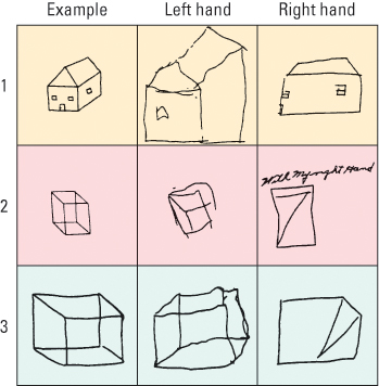
Other experiments with patients with split brains revealed that although the right hemisphere is unable to generate speech, it is much better than the left in solving visuospatial problems. When asked to arrange puzzle pieces to match a particular design or to copy three-dimensional pictures, each subject performed far better with the left hand (controlled by the right hemisphere) than with the right hand (controlled by the left hemisphere), even though all of the subjects were right-handed (see Figure 5.31). In more recent experiments, patients with split brains were asked to judge small differences between visual stimuli presented as pairs to either hemisphere. The results showed that the left hemisphere is as good as the right in judging nonspatial differences, such as differences in brightness or in color, but not as good as the right in judging spatial differences, such as differences in size or in the slant of two lines (Corballis, 2003).
How Patients with Split Brains Cope as Well as They Do
How do people who have had the split-brain operation get along in the world as well as they do? What keeps their two hemispheres from going off in opposite directions and creating continuous conflict between the two halves of the body? In some instances, especially shortly after the surgery, conflict does occur. One man described a situation in which, while he was dressing in the morning, his right hand was trying to pull his pants on while his left hand was trying to pull them back off (Gazzaniga, 1970); apparently, his right hemisphere wanted to go back to bed. But such conflicts are rare, and when they do occur, the left hemisphere (right hand) usually wins.
The patient’s ability to coordinate the two hemispheres probably involves several mechanisms. First, only the cerebral cortex and some parts of the limbic system are divided when the corpus callosum is cut. Motor centers that control movements of the larger muscles, such as those of the legs and arms (but not the fingers) lie in the lower, undivided parts, and some sensory information also passes from one hemisphere to the other by way of those lower routes (Reuter-Lorenz & Miller, 1998). The intact connections also apparently allow each hemisphere to inhibit the motor output of the other, so the more competent hemisphere can take control of any given task (Reuter-Lorenz & Miller, 1998). In addition, under normal conditions, when the eyes can move around and things can be felt with both hands, the two hemispheres can receive the same or similar information through their separate channels. Finally, each hemisphere apparently learns to communicate indirectly with the other by observing and responding to the behavior that the other produces, a process that Gazzaniga (1967) labeled cross-cueing. For example, the right hemisphere may perceive something unpleasant and precipitate a frown, and the left may feel the frown and say, “I’m displeased.”
183
Split-Brain Insight into Consciousness: The Left-Hemisphere Interpreter
30
How do studies of patients with split brains tend to confirm and extend an idea about the nature of consciousness that was developed long ago by Sigmund Freud?
One of the most fascinating findings from studies of patients with split brains is that they rarely express surprise or confusion concerning the seemingly contradictory actions that their two hemispheres generate. When asked to explain some behavior triggered by the right hemisphere, the person (speaking from the left hemisphere) usually generates quickly and confidently a seemingly logical (but often false) explanation.
For instance, in one experiment a patient with a split brain was presented simultaneously with a picture of a chicken claw shown to the left hemisphere and a picture of a snow scene shown to the right hemisphere (Gazzaniga, 2000). Then he was shown (in a way that could be seen by both hemispheres) a set of pictured objects and was asked to point with both hands to the one most closely related to what he had seen on the screen. He immediately pointed with his right hand to the chicken (clearly related to the chicken claw) and with his left hand to the snow shovel (clearly related to the snow scene). When asked why he was pointing to these two objects, his left, speaking hemisphere responded, “Oh, that’s simple. The chicken claw goes with the chicken, and you need a shovel to clean out the chicken shed.” He seemed completely satisfied by this explanation. In other experiments, Gazzaniga and his colleagues found that they could induce states of annoyance or of pleasure in patients with a split brain by flashing annoying or pleasant scenes to their right hemispheres. When asked to explain their feelings, the patients always came up with plausible (but clearly false) answers. For example, they might comment on some aspect of the experimental equipment—or of the experimenter’s behavior—that was either annoying them or pleasing them.
Such observations led Gazzaniga (2000) to posit that one of the natural functions of the left hemisphere is to interpret, or try to make logical sense of, everything that the person does. You might think of this left-hemisphere interpreter as analogous to the public relations department of a business or government. Its role is to tell stories, both to the self and to others, designed to make sense of the seemingly contradictory and irrational things that the person does. The idea of such an interpreter in the human brain or mind is by no means new. It was the centerpiece of a theory of consciousness proposed by Sigmund Freud (1912/1932) nearly 100 years ago. According to Freud, we do things because unconscious decision-making processes in our mind make us do them. But one part of our mind observes what we do and tells a running story about it; that story constitutes our conscious understanding of our actions and the reasons for them. The split-brain studies indicate that the neural mechanism for generating such stories is located in the left hemisphere and is intimately connected with the brain areas that generate speech.
Language Areas of the Left Hemisphere
Perhaps the most distinctively human behavioral ability is that of producing and understanding a complex, grammar-based language. Much of the left hemisphere of the human cortex is devoted in one way or another to language. Damage anywhere within large portions of the left hemisphere disrupts language ability, and the nature of the disruption depends on just where the destruction occurs.
184
Any loss of language ability resulting from brain damage is called aphasia [ǝ-fā´-zhiǝ]. Aphasias have been classified into a number of types, depending on the specific nature and degree of loss (Dronkers et al., 2000). The best known and most fully studied of these are two that were first described by nineteenth-century neurologists—one by Paul Broca and the other by Carl Wernicke.
Effects of Damage to Broca’s Area
31
What are the differences between Broca’s and Wernicke’s aphasias in (a) language production, (b) language comprehension, and (c) areas of the brain damaged?
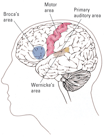
Broca (1861/1965) observed that people who had brain damage that included an area of the left frontal lobe now called Broca’s area, just anterior to the primary motor area (see Figure 5.32), suffered from a type of aphasia in which speech becomes labored and telegraphic, meaning that the minimum number of words are used to convey the message. The speech consists mostly of nouns and verbs, and a sentence is rarely more than three or four words long. If you ask a person with this disorder what he or she did today, the answer might be, “Buy bread store.” This disorder is now called Broca’s aphasia or, more descriptively, non-fluent aphasia. The lack of fluency in Broca’s aphasia suggests that neurons in Broca’s area are crucial for setting up the motor programs that are involved in fluent speech production. This conclusion is also consistent with the observation that Broca’s area lies very near to the region of the primary motor cortex that controls movements of the tongue and other speech muscles.
Research since Broca’s time indicates that people with damage to Broca’s area also have difficulty understanding language. They understand most of what they hear, but often fail to understand grammatically complex sentences. They become confused, for example, by sentences in which the agent and object of an action are reversed from the usual word order and the meaning cannot be inferred from the individual word meanings alone (Grodzinsky, 2000). Thus, they easily understand The boy pushed the girl (a simple, active sentence) or The apple was eaten by the boy (which can be understood just from word meanings because apples don’t eat boys), but The girl was pushed by the boy leaves them unsure as to who pushed whom. Research suggests that people with an intact Broca’s area understand that last sentence by transforming it mentally into its simpler, active equivalent—The boy pushed the girl. People lacking Broca’s area apparently fail to produce such transformations.
In sum, studies of people with damage to Broca’s area suggest that neurons in and around this area are crucial for at least two seemingly distinct language functions: (1) articulating words and sentences in a fluent manner and (2) transforming grammatically complex sentences that are heard into simpler ones in order to extract the meaning.
Effects of Damage to Wernicke’s Area
Wernicke (1874/1977) observed that people who had damage to a certain area of the left temporal lobe, now called Wernicke’s area (see Figure 5.32), suffered from a type of aphasia quite different from that described by Broca. These patients, unlike Broca’s aphasics, had difficulty understanding the meanings of words that they heard and also had difficulty finding the appropriate words to express the meanings that they wanted to convey.
The speech of patients with damage in Wernicke’s area is almost the opposite of that of Broca’s aphasics. It is rich in the little words that serve primarily to form the grammatical structure of a sentence—the articles (a, the), prepositions (such as of, on), and conjunctions (and, but). However, it is markedly deficient in the nouns, verbs, and adjectives that give a sentence its meaning. One such patient, asked to describe the contents of a simple picture, said: “Nothing the keesereez the, these are davereez and these and this one and these are living. This one’s right in and these are … uh … and that’s nothing, that’s nothing” (Schwartz, 1987). The inability to come up with the correct names of objects and actions leads to a heavy use of pronouns and nonsense words as substitutes. The speech retains its fluency and grammatical structure but loses its meaning.
185
This disorder is now called Wernicke’s aphasia, or, more descriptively, fluent aphasia. Theories to explain it generally center on the idea that neurons in and near Wernicke’s area are crucially involved in translating the sounds of words into their meanings and in locating, through connections to other cortical association areas, the words needed to express intended meanings (Dronkers et al., 2000). The location of this area near to the primary auditory area (see Figure 5.32) is consistent with the role of translating sounds into meaningful words (Geschwind, 1972).
Identifying Language Areas Through Neuroimaging
With PET or fMRI it is possible to determine which areas of the brain become more active when a person engages in a language-related task. Such studies tend in some ways to confirm the theories about the functions of Broca’s and Wernicke’s areas that were derived from studies of people with brain damage and in other ways to challenge those theories.
32
How was PET used to identify brain areas involved in word perception and production?
One of the earliest and most often cited of such studies was conducted by Steven Petersen and his colleagues (1989). These researchers used PET to image the brains of people as they carried out four types of language-related tasks that varied stepwise in level of complexity. At the first level (simplest task), the subjects simply gazed at a spot marked by crosshairs in the center of a video screen. At the second level, they continued to gaze at the crosshairs while they either saw (superimposed on the crosshairs) or heard (through earphones) a series of common English nouns. The third level was just like the second, except that now they were asked to speak aloud each word that they saw or heard. The fourth level was like the third, except that instead of simply repeating each noun, they were asked to think of and say aloud a verb that represented an action appropriate to the noun (for example, in response to hammer, they might say “pound”).
In order to identify the brain areas brought into play by each type of task, the researchers computed, for each small area of the brain, the difference between the average amount of activity during that task and the average amount during the task that ranked one level below that task. The results are depicted in Figure 5.33 and can be summarized as follows:
- Viewing or hearing words, without having to act on them in any way, resulted (as expected) in high activity in the relevant sensory areas—visual areas of the occipital lobe for viewing and auditory areas of the temporal lobe for hearing.
- Repeating aloud words that were seen or heard resulted in high activity in areas of the primary motor cortex that are involved in control of the vocal apparatus.
- Generating appropriate verbs in response to seen or heard nouns resulted in high activity in an area of the frontal lobe that encompassed Broca’s area and in a portion of the temporal lobe somewhat behind Wernicke’s area.
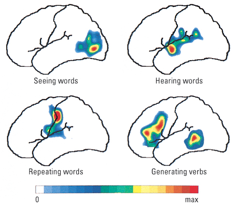
Notice that these results are not completely consistent with the theories about Broca’s and Wernicke’s areas developed from the brain-damage studies. Those theories would predict that Broca’s area should be involved in speaking words that have just been seen or heard as well as in speaking words that are mentally generated, and that Wernicke’s area, rather than the spot behind it, should be involved in generating words with the appropriate meaning. Neuroimaging studies are leading to new theories that implicate many cortical regions, not just Broca’s and Wernicke’s areas, in language comprehension and production (Bookheimer, 2002; Grodzinsky & Friederici, 2006).
186
SECTION REVIEW
The right and left hemispheres are specialized for different higher functions.
Split-Brain Studies
- Patients with split brains enable researchers to study the functioning of each hemisphere independent of the other.
- Results indicate that only the left hemisphere produces language in most people, while the right hemisphere is superior in visuospatial tasks.
- Split-brain studies also suggest that the verbal left hemisphere interprets a person’s own behavior, making “sense” of it even when the two hemispheres produce contradictory actions.
Language and the Left Hemisphere
- Much of the left hemisphere is devoted to language use.
- Aphasia is any language deficit due to brain damage. Deficits observed in Broca’s and Wernicke’s aphasia are used to infer functions of the corresponding brain areas.
- Results of neuroimaging studies are only partly consistent with aphasia research and have led to more extensive mapping of language areas in the brain.