Concept 4.2: Prokaryotic Cells Do Not Have a Nucleus
In terms of sheer numbers and diversity, prokaryotes are the most successful organisms on Earth. As we generalize about the features of these cells, bear in mind that there are vast numbers of prokaryotic species, and that the Bacteria and Archaea can be distinguished from one another in numerous ways. These differences, and the vast diversity of organisms in these two domains, are the subject of Chapter 19.
64
Prokaryotic cells, with diameters or lengths in the range of 1-10 μm, are generally smaller than eukaryotic cells, whose diameters or lengths are usually in the range of 10-100 μm. While some prokaryotes exist as single cells, other types form chains or small clusters of cells, and in some cases certain cells in a group perform specialized functions.
Prokaryotic cells share certain features
All prokaryotes have the same basic structure (FIGURE 4.5):
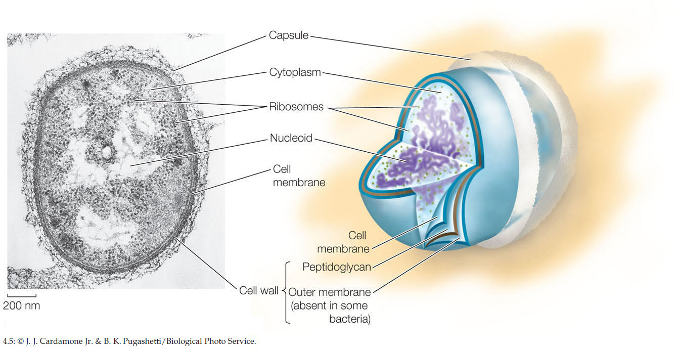
- The cell membrane encloses the cell, separating its interior from the external environment, and regulates the traffic of materials into and out of the cell.
- The nucleoid is a region in the cell where the DNA is located. DNA is the hereditary material that controls cell growth, maintenance, and reproduction (see Chapter 3).
- The rest of the material inside the cell is called the cytoplasm. The cytoplasm consists of a liquid component, the cytosol, and a variety of insoluble filaments and particles, the most abundant of which are ribosomes (see above).
- The cytosol consists mostly of water containing dissolved ions, small molecules, and soluble macromolecules such as proteins.
- Ribosomes are complexes of RNA and proteins that are about 25 nm in diameter. They can be visualized only with the electron microscope. They are the sites of protein synthesis, where the information encoded by nucleic acids directs the sequential linking of amino acids to form proteins. The cytoplasm is not a static region. Rather, the substances in this environment are in constant motion. For example, a typical protein moves around the entire cell within a minute, and it collides with many other molecules along the way. This constant motion helps ensure that biochemical reactions proceed at sufficient rates to meet the needs of the cell. Prokaryotes may look simple, but in reality they are functionally complex, carrying out thousands of biochemical reactions.
Specialized features are found in some prokaryotes
As they evolved, some prokaryotes developed specialized structures that gave them a selective advantage in their particular environments. These cells were better able to survive and reproduce than cells lacking the specialized structures.
Cell Walls
Most prokaryotes have a cell wall located outside the cell membrane. The rigidity of the cell wall supports the cell and determines its shape. The cell walls of most bacteria, but not those of archaea, contain peptidoglycan, a polymer of carbohydrates that is linked at regular intervals to short peptides. Cross-linking among these peptides results in a single giant molecule that surrounds the entire cell. In some bacteria, another layer, the outer membrane (a polysaccharide-rich phospholipid membrane), encloses the peptidoglycan layer (see Figure 4.5). Unlike the cell membrane, this outer membrane is relatively permeable, allowing the movement of molecules across it.
Enclosing the cell wall of some bacteria is a slimy layer composed mostly of polysaccharides, referred to as the capsule. In some cases these capsules protect the bacteria from attack by white blood cells in the animals they infect. Capsules also help keep the cells from drying out, and sometimes they help bacteria attach to other cells. Many prokaryotes produce no capsule, and those that do have capsules can survive even if they lose them, so the capsule is not essential to prokaryotic life. Some strains of the bacterium Streptococcus pneumoniae have a capsule and can cause pneumonia in humans and other mammals; however, non-encapsulated strains do not cause the disease.
65
Internal Membranes
Some groups of bacteria—including the cyanobacteria—carry out photosynthesis: they use energy from the sun to convert carbon dioxide and water into carbohydrates. These bacteria have an internal membrane system that contains molecules needed for photosynthesis. The development of photosynthesis, which requires membranes, was an important event in the early evolution of life on Earth. Other prokaryotes have internal membrane folds that are attached to the cell membrane. These folds may function in cell division or in various energy-releasing reactions.
A bacterium with enclosed compartments would have several evolutionary advantages. Chemicals could be concentrated within particular regions of the cell, allowing chemical reactions to proceed more efficiently. Certain biochemical activities could be segregated within compartments with more favorable conditions for those reactions, such as a different pH from the rest of the cell.
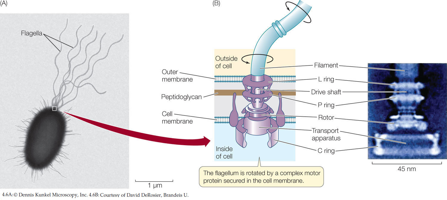
Flagella
Some cells swim by using appendages called flagella, which sometimes look like tiny corkscrews (FIGURE 4.6A). These movements are important in allowing the cells to swim toward food, for example. In bacteria, the filament of the flagellum is made of a protein called flagellin. A complex motor protein (see Concept 4.4) spins each flagellum on its axis like a propeller, driving the cell along. This motor protein is anchored to the cell membrane and, in some bacteria, to the outer membrane of the cell wall (FIGURE 4.6B). We know that flagella cause the motion of cells because if they are removed, the cells do not move. Flagellar motion can result in impressive speeds. The prokaryote Methanocaldococcus has been clocked at over 400 μm (500 body lengths) per second. For a car, this would mean 4,000 miles per hour.
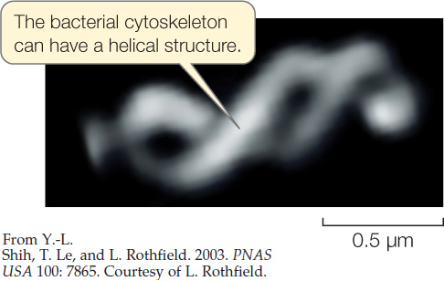
Cytoskeleton
The cytoskeleton is the collective name for filaments made up of polymers of monomer subunits that play roles in cell division or in maintaining the shapes of cells. One such protein forms helical structures that extend down the lengths of rod-shaped bacterial cells, helping maintain their shapes. This protein is similar to actin in eukaryotic cells, which we will discuss next.
CHECKpoint CONCEPT 4.2
- Compare the structures and functions of bacterial cell walls with those of bacterial cytoskeletons.
- What is the evolutionary advantage of bacteria that have flagella over bacteria that do not?
The prokaryotic cell is one of two broad types of cells recognized in cell biology. The other is the eukaryotic cell. Eukaryotic cells, and multicellular eukaryotic organisms, are more structurally and functionally complex than prokaryotic cells.