Concept 29.4: Animals Exhibit Division of Labor, but Each Cell Must Make Its Own ATP
Throughout the living world, structure and physiology—form and function—are intimately related. Often, form cannot be understood without understanding function, and vice versa.
Looking at the structure of animals, perhaps the first point worth noting is that animals typically consist mostly of water. Adult mammals (including us), for example, tend to be about 60 percent water. That water is said to be distributed in various body “compartments,” which by definition are location categories rather than specific organs. Typically, most of the fluid in an animal’s body is located within its cells. This fluid is called intracellular fluid, and the water in all the intracellular fluid throughout the body is said to be in the intracellular compartment. The rest of the fluid in the body is the extracellular fluid, said to be in the extracellular compartment. There are two subcategories of extracellular fluid, which we have already mentioned: the fluid portion of blood—termed the blood plasma—and the extracellular fluid found between cells in tissues throughout the body—termed the tissue fluid or, more formally, interstitial fluid.
Fluid compartments are separated from one another by physiologically active epithelia and cell membranes
How are the various fluid compartments kept separate in the body? There are two different scales of space we need to consider to answer this question, and they correspond to separation by epithelia and by cell membranes.
Epithelia
Consider the following three examples in which fluids of two types lie close together:
- Blood plasma inside a blood capillary and tissue fluid surrounding the outside of the capillary
- Watery fluid inside the lumen (central cavity) of a vertebrate’s intestines and tissue fluid in the intestinal wall
- Pond water on the outside of a submerged frog’s skin and tissue fluid in the tissue of the frog’s skin
In all three cases, the two fluids are separated by an epithelium (plural epithelia), which is a sheet of cells (epithelial cells) that covers a body surface or organ or lines a body cavity. The simplest and most common type of epithelium, called a simple epithelium, consists of just a single layer of cells that rests on a nonliving, highly permeable basement membrane (also called basal lamina; not to be confused with a cell membrane) that the cells help synthesize (FIGURE 29.13). Simple epithelia are exceedingly common. In our bodies, for example, a simple epithelium lines all our blood vessels and our intestines, as in the cases above. A simple epithelium also lines our kidney tubules, mammary glands, and sweat glands. In a frog, the skin consists entirely of living cells (unlike our skin, which has dead cells on the outside), and the outermost part of the skin is an epithelium.
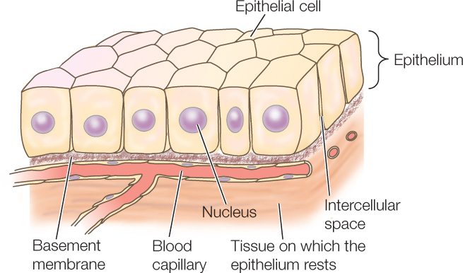
Besides helping compartmentalize the body by separating fluids, an epithelium typically has numerous functional capabilities and plays major physiological roles. The cells of an epithelium, for example, commonly pump ions between the fluids on either side of the epithelium. Some epithelia secrete substances such as hormones, mucus, digestive enzymes, milk, or sweat. Some absorb nutrients from the gut. Other epithelia serve sensory functions, including smell and taste.
616
Cell Membranes
At a much smaller scale, separation of fluid compartments is by cell membranes. Each cell has intracellular fluid inside separated from the extracellular fluid bathing the cell on the outside. The cell membrane separates the two. Cell membranes, like epithelia, have numerous functional capabilities and play major physiological roles. Cell membranes pump ions between the intracellular fluid and extracellular fluid. They often also play key roles in producing and receiving physiological signals.
LINK
You can review the properties of cell membranes and their transport and signaling functions in Chapter 5
Animals exhibit a high degree of division of labor
In multicellular organisms, individual cells need not perform for themselves all the functions required for life. Instead, cells can become specialized to perform just certain functions and depend on other cells in the body to perform others. Animals display a high degree of such division of labor.
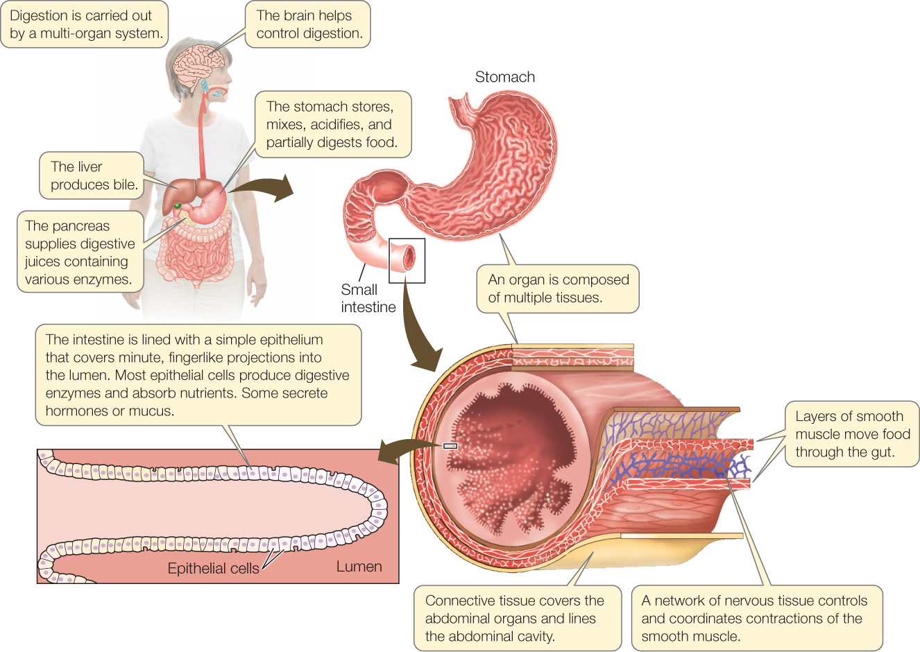
For understanding the structure and function of an animal’s body, we usually recognize cells, tissues, organs, and multi-organ systems as ever-more-complex levels in the hierarchy of organization of the animal body. A tissue is an assemblage of cells of similar type. An organ consists of two or more types of tissue with a defined structural relationship to each other. A multiorgan system consists of multiple organs working together.
As an example, FIGURE 29.14 shows parts of the human digestive tract, including the detailed structure of the beginning of the small intestine. The small intestine itself is an organ. It consists of several tissue types: muscle layers, nervous tissue, connective tissue, an epithelium lining the lumen (central cavity), and so forth. Each of these tissue types consists principally of a single, specialized type of cell, such as smooth muscle cells in the muscle layers and nerve cells in the nervous tissue. During its normal function, the small intestine receives partially digested food from the stomach, digestive juices from the pancreas, and an emulsifying agent called bile from the liver, and it is controlled in part by the brain. The intestine, stomach, pancreas, liver, and brain are all organs, and they work together as a multi-organ system to process a meal.
617
Each organ in an animal’s body is specialized to perform certain functions and is utterly dependent on other organs to perform other vital functions. The small intestine of a mammal, for example, requires O2 for its survival and thus has a life-and-death dependency on the lungs, which take up O2 from the atmosphere. The intestine also cannot carry out reproduction. Instead, an animal depends on its ovaries or testes to produce gametes. In turn, the lungs, ovaries, and testes depend on the intestine to supply them with nutrients.
Go to ACTIVITY 29.2 Tissues and Cell Types
PoL2e.com/ac29.2
Division of labor requires a rapid transport system
As a consequence of division of labor, organs often require inputs of materials from other organs, as exemplified by the small intestine needing O2 from the lungs. Organs also need materials they make to be delivered to other organs. For example, wastes need to go to the kidneys and hormones need to be delivered to their targets.
Because of these needs, a rapid transport system is typically required for division of labor to be successful. Most types of animals have a circulatory system that serves this role. In many cases, the need for O2 transport is the most pressing of all transport needs. For this reason, the sophistication of the circulatory system is usually correlated with metabolic rate (see Concept 32.1). Animals with high metabolic rates require high rates of O2 transport and have evolved circulatory systems with exceptional abilities for rapid O2 transport.
LINK
The diverse circulatory systems of animals are compared in Concepts 32.1 and 32.2
Each cell must make its own ATP
One of the most important principles in the study of animals is the fact that each cell must make its own ATP. ATP serves as the immediate energy source for most cellular functions, but it is not transported from cell to cell. Each cell must make its own ATP, and the rate of ATP supply in each cell depends on that particular cell’s ability to synthesize ATP.
How, then, is energy transported in an animal’s body? Energy is transported from organ to organ and from cell to cell in the form of food compounds, such as fatty acids or the sugar glucose, that can be broken down to release chemical energy. Each cell in the body takes up fatty acids or glucose from the blood and then breaks these compounds down internally to make ATP from the chemical energy released from chemical bonds.
Animal cells have aerobic and anaerobic processes for making ATP
Most animal cells make most of their ATP by processes that require O2. Such processes are termed aerobic, or oxygen-requiring. In aerobic ATP production, there is, speaking roughly, a fixed relationship between ATP production and O2 use. Thus the rate at which a cell can make ATP aerobically depends in part on the rate at which the cell is supplied with O2 by the circulatory system or by other O2-transport systems. Think back to the man running steadily on a treadmill in our opening photo. For a muscle cell in his legs to make ATP aerobically at a high rate—enabling the cell to have a high muscular power output—the cell must receive O2 at a high rate.
Aerobic ATP production takes place mostly in the mitochondria of a cell, by the processes of electron transport and oxidative phosphorylation. During aerobic ATP production, all types of food molecules—sugars, lipids, and proteins—can be used as fuels, that is, as sources of chemical energy for ATP synthesis.
Some cells also have processes for making ATP without O2. These mechanisms—termed anaerobic, meaning without oxygen—are not universally present. Brain cells in nearly all vertebrates, for example, have little ability to make ATP anaerobically. This explains why the vertebrate brain is quickly damaged by the absence of O2 (as, for example, during a heart attack or stroke). However, many types of cells—including muscle, kidney, and gut cells—have well-developed mechanisms for making ATP anaerobically.
LINK
Aerobic and anaerobic metabolism are discussed in Concepts 6.2 and 6.3, respectively, and are compared in Concept 33.3
The most common process of anaerobic ATP production in animals is anaerobic glycolysis. In this mechanism, glycolysis (see Figure 6.7) is the only ATP-producing biochemical pathway that is operational (glycolysis is not itself either aerobic or anaerobic, but it is termed anaerobic glycolysis when it takes place where O2 is lacking). The mitochondria, which require O2, do not participate in anaerobic glycolysis. With only glycolysis operational, only sugars can be used as fuels. Anaerobic glycolysis produces just a small amount of ATP per sugar molecule, but it can process sugar molecules at exceedingly high rates and thus make ATP at extraordinary rates. However, ATP cannot be made for very long because anaerobic glycolysis produces lactic acid as its final product (see Figure 6.12A), and the lactic acid accumulates in the animal’s body, eventually leading to imbalances that cause the process to stop. When people compete in mile or half-mile runs, much of the ATP used by their leg muscles is made anaerobically. This helps explain why the intensity of muscular work is high but the exercise cannot be continued for very long. Marathons must be run more slowly (mostly aerobically) to avoid this sort of fatigue.
618
CHECKpoint CONCEPT 29.4
- Could a single cell extracted from a multicellular organism survive on its own, similar to the way the lone cell of a unicellular organism does? Why or why not?
- In what form is energy transported from cell to cell?
- Why is permeability an essential attribute of compartmentalizing structures such as cell membranes and simple epithelia?
Before ending our introductory chapter on animals, we will now turn our spotlight on two additional topics of great importance: phenotypic plasticity and the ways animals control how they function.