Concept 33.2: Skeletal Muscles Pull on Skeletal Elements to Produce Useful Movements
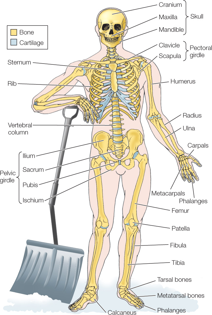
Skeletal systems provide rigid structural elements against which the skeletal muscles pull to create directed movements. Two major types of skeletal systems exist in animals. In vertebrates, the skeleton is surrounded by the other tissues of the body (FIGURE 33.8) and is called an endoskeleton (endo, “inside”) because it is deep inside the body. In arthropods, such as crabs and insects, the skeleton encases the rest of the body and is an exoskeleton (exo, “outside”).
In vertebrates, muscles pull on the bones of the endoskeleton
The endoskeleton consists mainly of bone in most adult vertebrates. Bone is composed principally of an extracellular matrix of collagen protein fibers with insoluble calcium phosphate crystals deposited among them, giving the bone hardness and rigidity. Bone also contains several types of living cells throughout life. The activities of these cells make bone a dynamic tissue. It is not inert, as many people tend to assume. Bone is remodeled—that is, repaired or altered in shape—throughout life by the activities of its living cells. Because of this process, a bone can undergo modest changes in shape or structure to adjust to the forces placed on it. Bone also serves as a reservoir of calcium for the rest of the body, being in dynamic exchange with soluble calcium in the extracellular body fluids. This exchange is under the control of hormones, such as parathyroid hormone (see Figure 35.10), and vitamin D. The endoskeleton also includes cartilage in its structure. Cartilage is a flexible, rather than hard, form of skeletal tissue that plays several roles, such as imparting flexibility to skeletal elements (e.g., the rib cage).
689
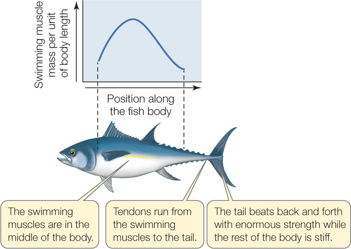
To produce movements, muscles and bones work together around joints, the structures where two or more bones come together. Muscles attach to bones by bands of flexible connective tissue called tendons. Tendons often extend across joints. In such cases, muscles on one side of a joint exert forces on bones on the opposite side of the joint. Tunas, which are among the most powerful swimmers of all fish, provide dramatic examples. A tuna swims by beating its tail fin, but there are no muscles of any size in its tail. The massive swimming muscles of a tuna are in the middle of its body (FIGURE 33.9). Forces developed by these muscles are transmitted to the tail by tendons that run from the swimming muscles to the tail fin across the joint at the base of the tail.
A muscle can exert force in only one direction. Thus for the bones at a joint to move in opposite directions, two muscles are required. These muscles work as an antagonistic pair in which the two have opposite actions: when one contracts, the other relaxes. You can see this action in the motions of your forearm (Figure 33.10A). When your biceps contracts, your triceps relaxes, and your forearm moves up because the tendons of the biceps cross the elbow joint to attach to the forearm. When your triceps contracts, your biceps relaxes, and your forearm moves down.
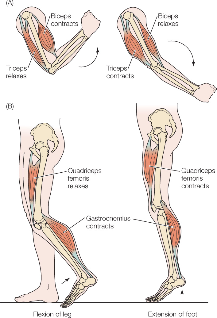
Different sets of muscles, coordinated by the nervous system, typically must work together to control complex sets of movements. For example, consider the large muscle on the back of your calf, the gastrocnemius muscle. When it contracts, the movement created depends on the state of contraction of the muscle on the front of your thigh, the quadriceps femoris (Figure 33.10B). If your quadriceps is not contracting strongly, contraction of your gastrocnemius bends your knee joint and lifts your calf as in walking. If your quadriceps is contracting strongly—an action that keeps your knee joint from bending—contraction of your gastrocnemius extends your foot as in jumping.
In arthropods, muscles pull on interior extensions of the exoskeleton
Crabs, lobsters, insects, and other arthropods have an exoskeleton that covers every part of their body. It is composed of the polymerized carbohydrate chitin, and in crabs and lobsters it is extensively hardened by deposition of calcium-containing minerals. An exoskeleton functions like armor, protecting the soft tissues of the body inside. However, the exoskeleton of an arthropod cannot grow after it is formed. As an arthropod grows, it must therefore replace its exoskeleton periodically with a new, larger version.
690
LINK
The process of shedding an exoskeleton—called molting—is controlled by hormones, as described in Concept 35.5
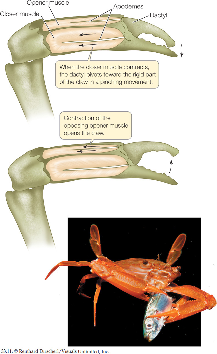
The skeletal muscles have a different relationship to the skeleton in arthropods than they do in animals with endoskeletons. Typically, where there is a joint between two parts of the exoskeleton, the exoskeleton projects inward, forming structures called apodemes to which the muscles inside the body attach. The large “pinching” claws at the end of a crab’s front legs provide an excellent illustration. One side of each claw does not move, but the other side (the dactyl) connects to the leg by a joint and is moveable. At the joint, the moveable element of the claw has two apodemes to which two muscles attach (FIGURE 33.11). These muscles act as an antagonistic pair. When the larger of the two contracts, it pulls the claw closed with great force (leading to a pinching movement and a loud scream if your finger is caught!). The smaller of the two opens the claw with less force when it contracts.
LINK
The diverse structures and compositions of exoskeletons are detailed in Concept 23.3 and Concept 23.4
Hydrostatic skeletons have important relationships with muscle
An animal is said to have a hydrostatic skeleton if its body or a part of its body becomes stiff and skeleton-like because of a high fluid pressure inside. The most familiar hydrostatic skeleton is the human erected penis; blood pressure, produced by the beating of the heart muscle, gives it its rigid, skeleton-like character (see Concept 37.2).
691
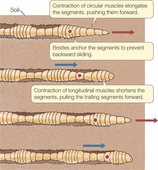
An earthworm digs through soil by using muscles in its body wall to create a hydrostatic skeleton (FIGURE 33.12). Each time the worm moves forward (red arrows in the figure), contractions of certain muscles raise the pressure inside the worm to a high enough level that the front end of the worm’s body expands forward through the soil, much as a ribbon-shaped balloon expands when inflated. The worm then anchors its front end and pulls its back end toward its front end (blue arrows in the figure) before repeating the process. Clams do much the same thing to dig into sand. Through muscular contractions, they cyclically inflate their foot under pressure, so that the foot shoves deeper into the sand, and then pull the rest of their body down toward the foot.
CHECKpoint CONCEPT 33.2
- What are some biological structures, apart from muscle, that allow animals with endoskeletons to perform complex movements?
- Can endoskeletal components continue to change shape and structure after an animal is done growing?
- Are all muscles that function in locomotion necessarily attached to hard skeletal elements?
Now that we have looked at how skeletal muscles work together with different skeletal systems, let’s turn to the factors that affect the performance of the skeletal muscles.