Concept 35.3: The Vertebrate Hypothalamus and Pituitary Gland Link the Nervous and Endocrine Systems
The pituitary gland, found at the base of the brain (FIGURE 35.6A; see also Figure 34.25), is sometimes called the “master gland” because it secretes hormones that control the functions of many other glands. It is attached by a stalk to a part of the brain called the hypothalamus. The pituitary has two parts—the anterior pituitary (adenohypophysis) and posterior pituitary (neurohypophysis). The two have different developmental origins, which helps account for marked differences in the way they function. Both parts of the pituitary have close functional links with the brain.
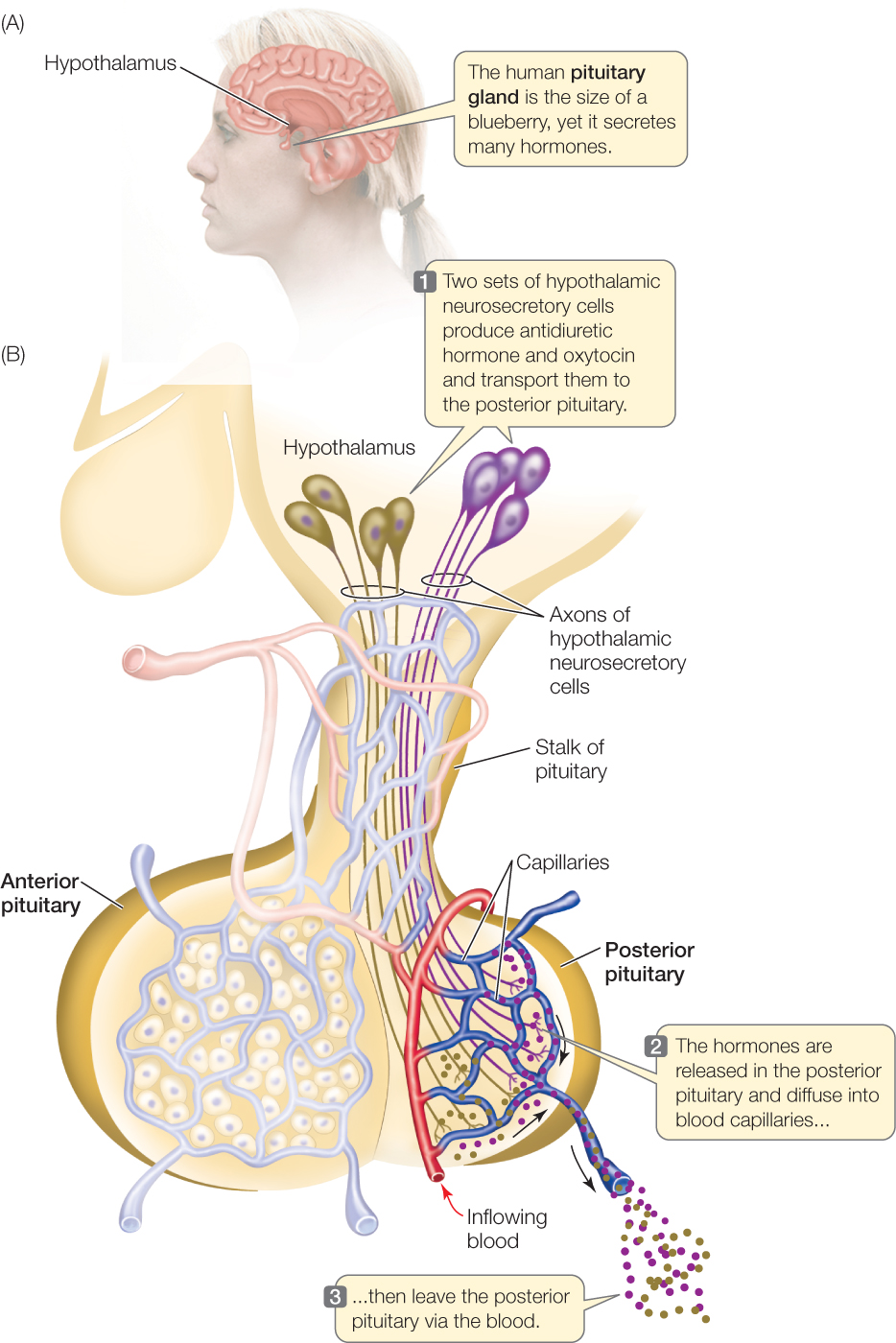
Hypothalamic neurosecretory cells produce the posterior pituitary hormones
The posterior pituitary is basically an extension of the brain (FIGURE 35.6B). Its hormones are synthesized and secreted by brain neurosecretory cells. The cell bodies of these cells are in the hypothalamus, where they receive synapses from other, ordinary brain cells (neurons). The axons of the neurosecretory cells extend into the posterior pituitary, where they terminate in a rich blood capillary network, forming a neurohemal organ where the hormones of the neurosecretory cells are secreted into the blood.
An important general principle is that different sets of neurosecretory cells secrete different hormones. We can apply this principle as we focus on the hypothalamus and posterior pituitary. In most mammals and also in many other vertebrates, there are two sets of neurosecretory cells, which secrete two hormones, in the posterior pituitary system. Both hormones are short peptides containing nine amino acid units. In mammals these two hormones are antiduiretic hormone (ADH; also called vasopressin) and oxytocin. ADH helps control water excretion by the kidneys, as we will discuss in Chapter 36. Oxytocin controls milk ejection from the mammary glands and helps control contractions of the uterus during the birth process (see Figure 29.17).
741
Signals produced by the brain control secretion by these neurosecretory cells. In this way, the enormous computational and integrative powers of the brain can be marshaled to control secretion of the posterior pituitary hormones. When activated, the neurosecretory cells generate action potentials that travel to their axon terminals, leading to hormone release into the blood.
In short, in the posterior pituitary system, brain neurons control the secretion of hormones into the general circulation by neurosecretory cells. This is one type of interface between the nervous and endocrine systems—a type of interface that we commonly find in both vertebrates and invertebrates.
Hormones from hypothalamic neurosecretory cells control production of the anterior pituitary hormones
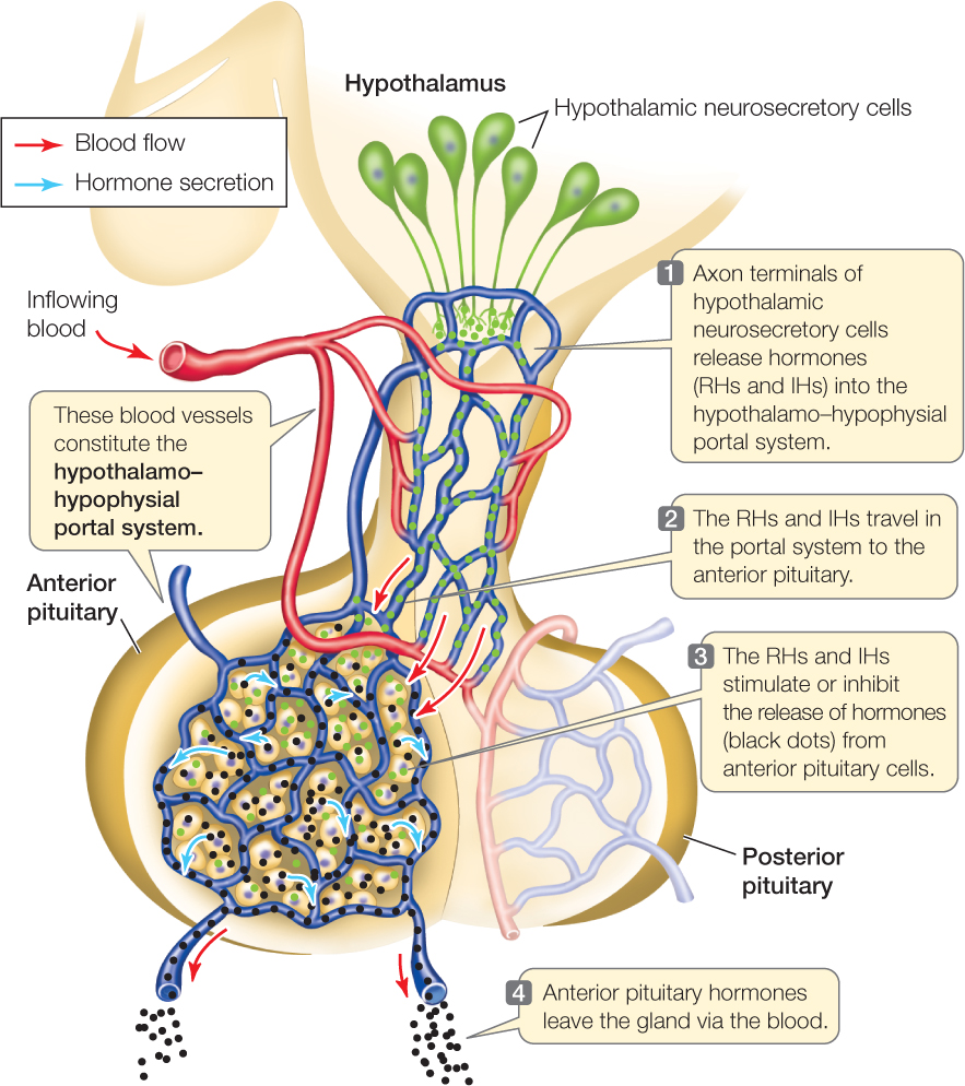
Unlike the posterior pituitary (which is a neurohemal organ), the anterior pituitary is an endocrine gland. The anterior pituitary hormones are synthesized in the anterior pituitary gland itself by nonneural endocrine cells (FIGURE 35.7). All the hormones are peptides or closely related to peptides.
Four of the hormones are known as tropins, or tropic hormones, meaning their principal functions are to control the activities of other endocrine glands. They are adrenocorticotropic hormone (ACTH), follicle-stimulating hormone (FSH), luteinizing hormone (LH), and thyroid-stimulating hormone (TSH). A different set of pituitary cells produces each tropic hormone. ACTH helps control the adrenal cortex (see Figure 35.5). Both FSH and LH, which together are called gonadotropins, control the gonads: the testes in males and the ovaries in females. TSH controls the thyroid gland.
The anterior pituitary gland also produces several other hormones. These hormones act on non-endocrine targets and therefore are not tropic hormones. They include growth hormone (GH; see Figure 35.4A), which acts on a wide variety of tissues to promote growth. They also include prolactin (which we’ve already mentioned), and melanocyte-stimulating hormone, which is named for its action on skin-color cells in amphibians.
How is secretion of the anterior pituitary hormones controlled? A critical role is played by a specialized vascular network, the hypothalamo–hypophysial portal system (“hypophysis” is an alternative name for pituitary). This vascular system consists of two capillary beds that are connected so that blood flows through them sequentially. Blood flows first through a capillary bed in the hypothalamus. Neurosecretory cells in the hypothalamus secrete hormones into the blood in this capillary bed. When the blood flows out of the hypothalamus, it travels a short distance to the second capillary bed, which is in the anterior pituitary. In this way, the hormones secreted by the hypothalamic neurosecretory cells are brought to the pituitary nonneural endocrine cells. These hormones control secretion by the endocrine cells in the pituitary. A typical sequence of action begins when ordinary brain neurons that synapse on the hypothalamic neurosecretory cells stimulate those cells to secrete hormones. Those hormones then travel to the anterior pituitary and control secretion by the anterior pituitary nonneural endocrine cells.
742
The hormones added to the blood by the neurosecretory cells in the hypothalamus are called releasing hormones (RHs) and inhibiting hormones (IHs; also called release-inhibiting hormones). Each RH or IH is specific in its actions. For example, there are separate RHs for growth hormone and thyroid-stimulating hormone. When the RH for growth hormone is secreted in the hypothalamus, it affects the specific cells in the anterior pituitary that secrete growth hormone, stimulating them to secrete (release) their hormone into the general circulation. The RH for thyroid-stimulating hormone, in contrast, stimulates the specific pituitary cells that secrete TSH.
In summary, in the anterior pituitary system, neurosecretory cells in the brain control the secretion of hormones into the general circulation by other endocrine cells. This is a second type of interface between the nervous and endocrine systems. It too is a type of interface that we commonly find in both vertebrates and invertebrates.
Endocrine cells are organized into control axes
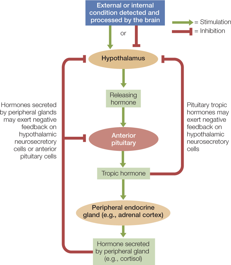
A typical sequence in vertebrates is for a hormone secreted by brain neurosecretory cells to control the secretion of a tropic hormone by anterior pituitary cells, and for the tropic hormone then to control hormone secretion by the cells in a gland elsewhere in the body. A system of this type—in which endocrine cells act on each other in sequence—is called an axis.
One example is the hypothalamus–pituitary–adrenal cortex (HPA) axis. Some of the steroid hormones secreted by the adrenal cortex, including a class called glucocorticoids, are important in responding to long-term stress. When the brain, using its great computational capacity to integrate inputs, detects that an animal is under stress, some of the cells in the hypothalamus secrete corticotropin-releasing hormone. This releasing hormone hor call term cap der cort travels in the portal system to cells in the anterior pituitary gland that respond by increasing secretion of the tropic hormone adrenocorticotropic hormone (ACTH). ACTH then travels in the general circulation to the adrenal cortex, which responds by increasing secretion of glucocorticoids into the blood. In primates, the principal glucocorticoid is cortisol (see Figure 35.4B).
743
Negative feedback often takes place within an axis. How does it work? One way is that the hypothalamic neurosecretory cells and the endocrine cells of the anterior pituitary are often under negative-feedback control by the hormones of their target glands (FIGURE 35.8). For example, when the blood concentration of cortisol (or other glucocorticoids) increases following stimulation of the adrenal cortex by the HPA axis, blood cortisol tends to cause cells in the hypothalamus to reduce release of corticotropin-releasing hormone and cells in the anterior pituitary to reduce secretion of ACTH. This negative feedback helps stabilize the blood cortisol concentration at a relatively constant level.

Go to ANIMATED TUTORIAL 35.1 The Hypothalamus and Negative Feedback
PoL2e.com/at35.1
Hypothalamic and anterior pituitary hormones are often secreted in pulses
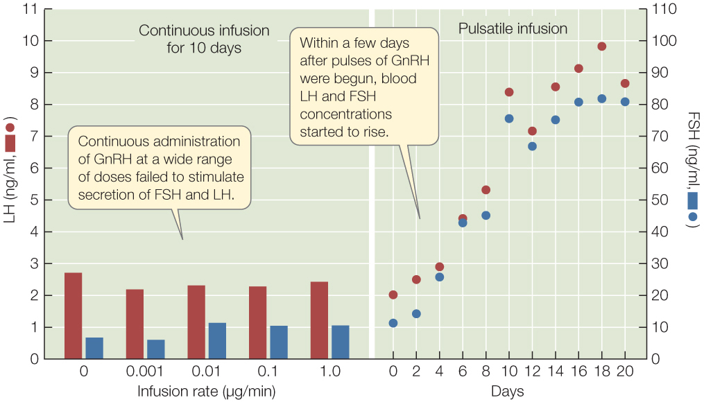
In many cases the brain stimulates the neurosecretory cells in the hypothalamus to secrete in pulses instead of continuously. Corticotropin-releasing hormone, for example, is typically secreted in mammals in two or three pulses per hour. Pulsed secretion of RHs or IHs often proves to be necessary for these hormones to act (FIGURE 35.9). Continuous secretion can lead target cells in the pituitary to reduce their numbers of RH or IH receptor molecules and thus reduce their sensitivity. Pulsed secretion is hypothesized to prevent this loss of sensitivity. At the next step in the axis, the anterior pituitary hormones are often also secreted in pulses.
Investigation
HYPOTHESIS
The effectiveness of GnRH depends on the dose, not on the pattern of delivery.
METHOD
- Administer GnRH to rhesus monkeys in which the hypothalamic cells responsible for GnRH secretion have been destroyed. Administer GnRH either continuously at 0.001, 0.01, 0.1, or 1.0 μg/min, or in pulses (one 6-min pulse per hr) at 1.0 μg/min.
- Measure the concentrations of LH and FSH in the blood.
RESULTS
CONCLUSION
The hypothesis is not supported: the effectiveness of the GnRH depends on the pattern of delivery.
ANALYZE THE DATA
- For delivery in pulses, GnRH was administered at 1 μg/min for 6 minutes once per hour. Which continuous rate delivered the same amount of GnRH per hour as the delivery in pulses? How much GnRH was delivered per hour at the lowest continuous rate? How much lower was this than the dose from pulses?
- For interpreting this experiment, why is it important that one of the continuous delivery rates provides the same total dose per hour as the pulsatile delivery?
- We now know that the anterior pituitary stops responding to GnRH if GnRH is always present. What does this tell you about how fast each pulse of GnRH disappeared from the bloodstream? Be as quantitative as possible, taking account of the GnRH dose that you calculated for the lowest continuous rate above.
Go to LaunchPad for discussion and relevant links for all INVESTIGATION figures.
aP. E. Belchetz et al. 1978. Science 202: 631–633.
744
CHECKpoint CONCEPT 35.3
- Considering the hierarchy of control, why might it be misleading to call the pituitary gland the “master gland”?
- Why are many anterior pituitary hormones called tropic hormones?
- Neurosecretory cells in the hypothalamus and cells in the anterior pituitary gland both have cortisol receptors. If they were to lose their cortisol receptors, these cells would immediately begin secreting unusually large amounts of corticotropin-releasing hormone and ACTH. Why?
The hypothalamus and anterior pituitary gland control the secretion of hormones from several other endocrine glands. We will now look at the hormones that some of these other glands release, and the functions of these hormones.