35.2 NEURON STRUCTURE
We have seen that nerve cells can be classified as sensory neurons, interneurons, or motor neurons. All share the same basic organization, adapted to their common function in receiving and transmitting information. Neurons have extensions that receive information and relay it to the cell body. Other extensions transmit information away from the cell body. Although all neurons share this basic plan, they differ remarkably in size and number of extensions, according to their specific function.
35-5
35.2.1 Neurons share a common organization.
Neurons all have some basic features in common. These include a large cell body from which emerge two kinds of fiberlike extension, the dendrites and axons (Fig. 35.4). These extensions are the input and output ends of the nerve cell. Both types of cellular extension can be highly branched, enabling neurons to communicate over large distances with many other cells. A neuron’s dendrites receive signals from other nerve cells or, in the case of sensory nerves, from specialized sensory endings. These signals travel along the dendrites to the neuron’s cell body. At the junction of the cell body and its axon, the axon hillock, the signals are summed. If the sum of the signals is high enough, the neuron fires an action potential, or nerve impulse, that travels down the axon. An action potential is a brief electrical signal transmitted from the cell body along one or more axon branches.
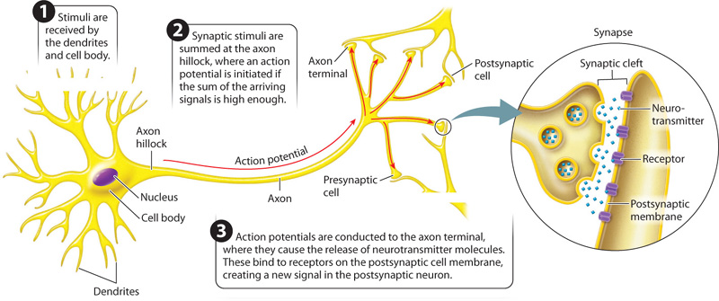
Axons generally transmit signals away from the nerve’s cell body. The end of each axon forms a swelling called the axon terminal. An axon terminal communicates with a neighboring cell through a junction called a synapse. A space, the synaptic cleft, separates the end of the axon of the presynaptic cell and the neighboring postsynaptic cell. The synaptic cleft is commonly only about 10 to 20 nm wide.
How does a signal cross the synaptic cleft? Molecules called neurotransmitters convey the signal from the end of the axon to the post-synaptic target cell. The arrival of a nerve signal at the axon terminal triggers the release of neurotransmitter molecules from vesicles located in the terminal. The vesicles fuse with the axon’s membrane, releasing neurotransmitter molecules into the synaptic cleft. The molecules diffuse across the synapse and bind to receptors on the plasma membrane of the target cell. The binding of neurotransmitters to these receptors causes a change in the electrical charge across the membrane of the receiving postsynaptic cell, continuing the signal in the second cell. Most neurons communicate by hundreds to thousands of synapses that they form with other cells. The massive number of interconnections enables the formation of complex nerve cell circuits and the transmission of information from one circuit to others in the nervous system.
Some neurons do not synapse with other neurons, but instead with other types of cell that produce some physiological response in the animal. Examples are muscle cells, which contract in response to nerve signals, and secretory cells in glands, which release hormones in response to nerve signals.
Question Quick Check 1
MK0k0a8OI021GvslR8ysXPnqv/Q7MjdRgQEtlYgkdZZLRlqvo5J2GZrBGHr0EwxyyIMX0hdzcmUGs8OA0vUPzLYWH1RIhOdM4VgkSXN2S9URNvW1jizskxXaybk3NlCj8rBOlG3HswERZUxw35.2.2 Neurons differ in size and shape.
In spite of their common organization, neurons show a remarkable diversity in size and shape. Some neurons, such as interneurons, are very short, whereas others, like the motor neuron that extends from your spinal cord to your big toe, are several feet long. The position of the cell body can also vary in different neurons (Fig. 35.5). Some sensory neurons involved in smell, sight, and taste have just two extensions coming off the cell body (Fig. 35.5a), whereas other neurons have many more (Fig. 35.5b and 35.5c).

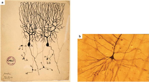
The cell bodies of some neurons have distinctive shapes. For example, pyramidal neurons, as their name suggests, have triangular-shaped cell bodies (Fig. 35.6). These cells, located in the mammalian brain, were elegantly drawn by the Spanish neuroanatomist Santiago Ramón y Cajal, who shared the 1906 Nobel Prize in Medicine or Physiology with the Italian scientist Camillo Golgi “in recognition of their work on the structure of the nervous system.”
35-6
The degree of branching of a neuron’s extensions can vary from one neuron to the next. A neuron’s dendrites form a finely branched field that receives inputs from many other nerve cells or receptors. A neuron with more dendritic branches receives inputs from more sources than one with fewer branches. Some neurons receive information only from nearby regions through a few dendrites that branch close to the cell body. Other neurons receive information from many branching dendrites and transmit their output along a single axon branch or only a few axon branches. Pyramidal cells provide an example (Fig. 35.6). They have many highly branched dendrites that converge on the cell body, but just a single axon. Pyramidal cells illustrate a general organizational feature of nervous systems: Information arriving from many sources often converges on a smaller subset of neurons.
Axons typically have fewer branches than dendrites. Signals from all the dendrites, converging on the neuron cell body, determine the strength, or frequency, and timing of signals carried by the neuron’s axon. The process of bringing together information gathered from different sources is referred to as the integration of information. The degree of branching and the number of synapses that a neuron makes with neighboring nerve cells, therefore, reflect how information is processed and integrated by the cell as part of a larger circuit.
35.2.3 Neurons are supported by other types of cell.
Neurons in many body regions, and in particular within the brain, are supported by other types of cell that do not themselves transmit electrical signals. Glial cells are a major class of supporting cell. Indeed, the human brain has more glial cells than neurons. Glial cells surround neurons and provide them with nutrition and physical support. During development, glial cells help orient neurons as they develop their connections.
Star-shaped glial cells called astrocytes contribute to the blood–brain barrier, a set of structural adaptations of the blood vessels supplying the brain that prevent pathogens and toxic compounds in the blood from entering the brain. Astrocytes surround blood vessels in the brain, limiting the size of compounds that can diffuse from the blood into the brain. Because nerve cells have limited capacity to regenerate after being damaged, protection of the brain and spinal cord is critically important. Nevertheless, lipid-soluble compounds such as alcohol, certain anesthetics, and other drugs readily diffuse across the blood–brain barrier. As a result, these compounds affect the functioning of the brain and an animal’s mental state. They can also lead to damage and even destruction of neurons in the brain.
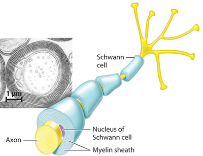
Glial cells also provide electrical insulation to vertebrate neurons. Glial cells form multiple lipid-rich layers or sheaths, called myelin, that wrap around the axons of neurons (Fig. 35.7). Myelin gives many nerves their glistening white appearance. Glial cells called Schwann cells insulate sensory and motor neurons, and glial cells called oligodendrocytes insulate cells in the brain and spinal cord. By insulating the axon, myelin greatly increases the speed of nerve signal transmission. As a result, the nervous system can respond rapidly to stimuli, as we will see after first discussing how nerve cells transmit electrical signals.