36.5 BRAIN ORGANIZATION AND FUNCTION
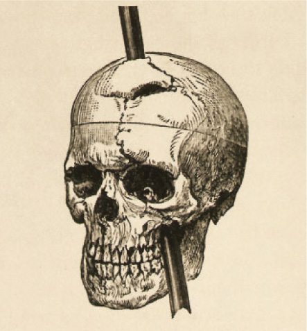
Up to this point, we have looked at how sensory information, such as smell, taste, hearing, and vision, is detected by sensory receptors. In each case, the sensory information is sent to the brain, where it is processed and integrated. Much of what we know about how the brain functions has been accumulated over many years by studies of patients who suffered brain injuries. A well-known example is Phineas Gage, a railroad worker who in 1848 survived severe damage to his brain’s frontal lobe after a tamping iron he was using to set a dynamite charge caused the dynamite accidentally to explode, sending the iron through his skull and the frontal cortex of his brain (Fig. 36.21). Gage had been hard-working, responsible, and well liked; after the accident he was given to fitful arguments, lacked self-control, and showed reduced intellectual capacity. These changes provided some of the first evidence that particular brain regions control particular aspects of a person’s behavior and cognitive function. The remainder of this chapter explores the functional organization of the vertebrate brain, highlighting examples that show how the brain integrates, organizes, and maps sensory information, and how memories of sights and sounds, for example, are formed that contribute to learning.
36.5.1 The brain processes and integrates information received from different sensory systems.
With the evolution of greater cognitive ability, animal brains became larger and more complex. Different brain regions became specialized to carry out different functions. The vertebrate brain is organized into a hindbrain, midbrain, and forebrain, with a cerebral cortex formed from a portion of the forebrain (Fig. 36.22a). The hindbrain and midbrain control basic body functions and behaviors, and the forebrain, particularly the cerebral cortex, governs more-advanced cognitive functions. The cerebral cortex of mammals—particularly primates and humans—is greatly expanded relative to that of other vertebrates.
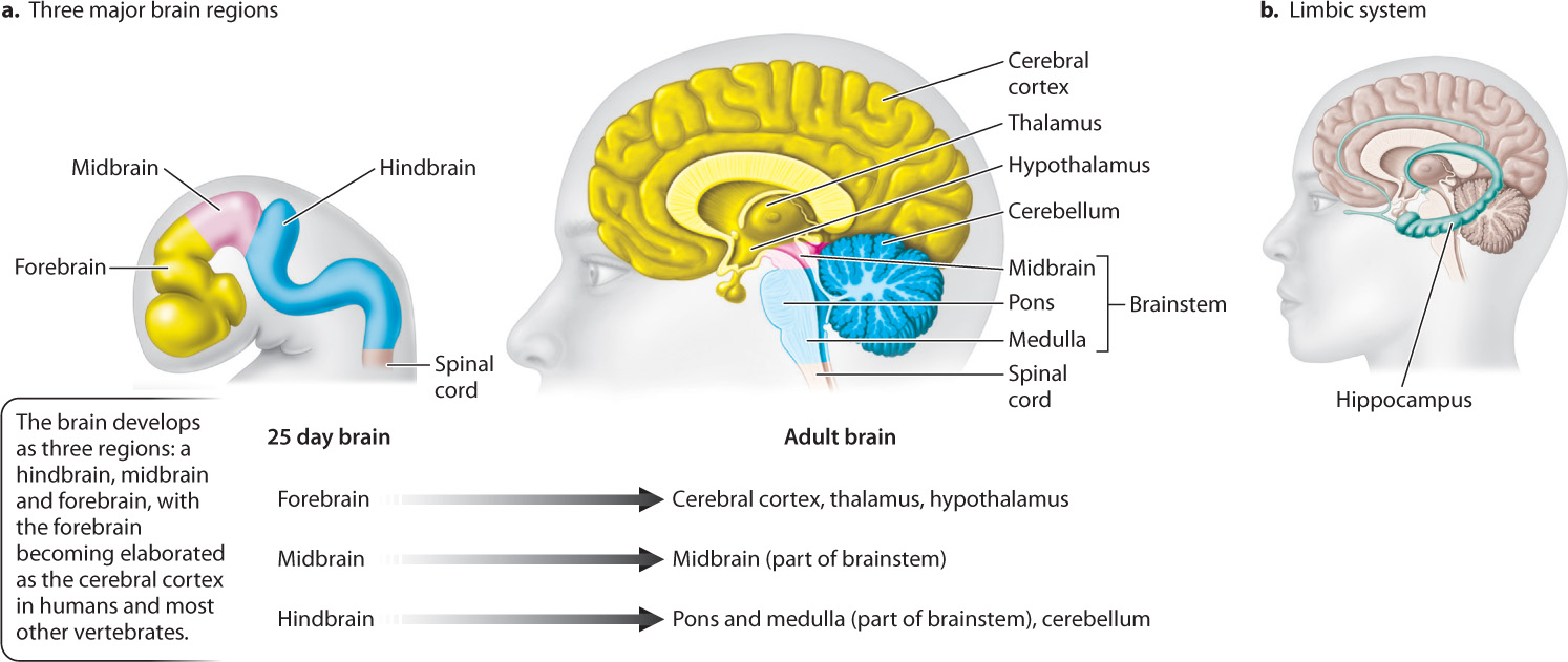
In adult vertebrates, the hindbrain develops into the cerebellum and a portion of the brainstem (Fig. 36.22a). The remainder of the brainstem develops from the midbrain. The cerebellum coordinates complex motor tasks, such as catching a ball or learning to write and talk. It integrates both motor and sensory information. The brainstem, consisting of the medulla, pons, and midbrain, initiates and regulates motor functions such as walking and controlling posture and coordinates breathing and swallowing. The brainstem activates the forebrain by relaying information from lower spinal levels. High levels of activity within the brainstem maintain a wakeful state; low levels enable sleep. If the midbrain is damaged, loss of consciousness and then coma result.
36-18
The forebrain consists of an inner brain region that forms the thalamus and the underlying hypothalamus, and a more anterior region that develops into the cerebrum, the outer left and right hemispheres of the cerebral cortex (Fig. 36.22a). The thalamus is a central relay station for sensory information sent to higher brain centers of the cerebrum. The hypothalamus interacts closely with the autonomic and endocrine systems to regulate the general physiological state of the body. In humans and most other primates, the cerebral cortex is the largest part of the brain, overseeing sensory perception, memory, and learning. Whereas the forebrain is essential to normal behavior in mammals, it appears less important for other vertebrates.
Other inner components of the forebrain constitute the limbic system, which controls physiological drives, instincts, emotions, and motivation and, through interactions with midbrain regions, the sense of reward (Fig. 36.22b). Stimulation of the limbic system can induce strong sensations of pleasure, pain, or rage, reinforcing learning related to basic physiological drives. A posterior region of the limbic system, the hippocampus, is involved in long-term memory formation, discussed in the next section.
Sensory information reaches the cerebral cortex from the cranial nerves and nerves passing through the spinal cord. This information passes through the brainstem, and then through the thalamus, the central relay station for sensory information. From the thalamus, information from each of the senses goes to a different region of the brain specialized to further process that information, in a manner discussed next.
36.5.2 The brain is divided into lobes with specialized functions.
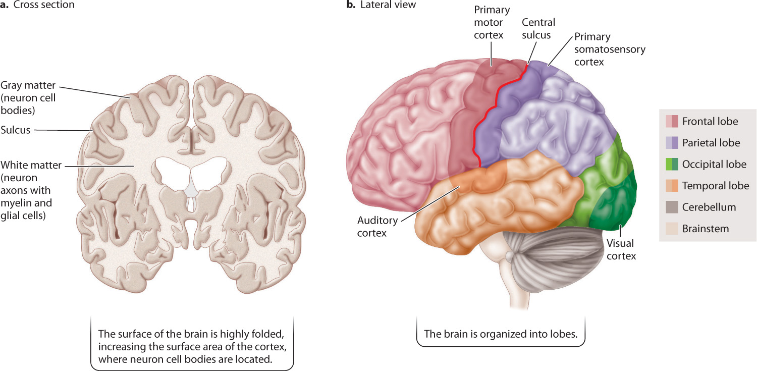
The cerebral hemispheres are the largest structures of the mammalian brain (Fig. 36.23). They consist of a highly folded outer layer of gray matter about 4 mm thick, called the cortex, that is made up of densely packed neuron cell bodies and their dendrites (Fig. 36.23a). The folds greatly increase the surface area of the cortex and are the result of selection for an increased number of cortical neurons within the limited volume of the skull. Deep inside the cortex is the white matter, which contains the axons of cortical neurons. It is the fatty myelin produced by glial cells surrounding the axon that makes this region of the brain white. These axons are the means by which neurons in different regions of the gray matter communicate with neurons in other regions of the cortex and in deeper regions of the forebrain, midbrain, and hindbrain.
36-19
Major regions of the cerebral hemispheres are defined by clearly visible anatomical lobes separated in most cases by deep crevices called sulci (singular, sulcus) (Fig. 36.23b). The frontal lobe is located in the anterior region of the cerebral cortex (behind your forehead). It is important in decision making and planning. The two parietal lobes are located posterior to the frontal lobe. The parietal lobes are separated from the frontal lobe by the central sulcus. The parietal lobe controls body awareness and the ability to perform complex tasks, such as dressing or brushing one’s hair. The two temporal lobes lie to the side, below the parietal lobes. They are involved in processing sound, as well as performing other functions discussed below. Located behind the parietal lobe, at the back of the brain, is the occipital lobe, which processes visual information from the eyes.
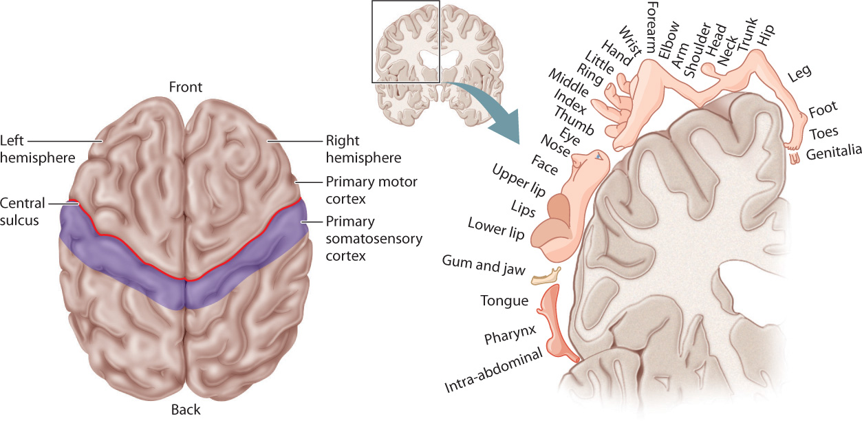
The central sulcus separates the primary motor cortex of the frontal lobe from the primary somatosensory cortex of the parietal lobe (Fig. 36.24). “Command” neurons in the primary motor cortex produce complex coordinated behaviors by controlling skeletal muscle movements. The primary somatosensory cortex integrates tactile information from specific body regions and relays this information to the motor cortex. Both cortices make connections to the opposite side of the body by sending axons that cross over in the brainstem and spinal cord. As a result, the right cortex controls the left side of the body and the left cortex controls the right side of the body.
36.5.3 Information is topographically mapped into the vertebrate cerebral cortex.
The primary motor cortex and somatosensory cortex are organized as maps that represent different body regions, as shown in Fig. 36.24. The neurons that control movements of the foot and human lower limb are located near the mid-axis of the motor cortex. Those that control motions of the face, jaws, and lips map to the side and farther down in the primary motor cortex. There is a similar topographic map in the somatosensory cortex for pressure and touch sensation. Regions such as the fingers, hands, and face that are involved in fine motor movements and distinguishing fine sensations are represented by larger areas of the cortex.
The auditory cortex in the temporal lobe is similarly organized as a map. This region processes sound information transmitted from the cochlea of the ear. Neurons in the auditory cortex are organized by pitch: Neurons sensitive to low frequencies are located at one end and neurons sensitive to high frequencies at the other, helping to distinguish fine differences in pitch. With the evolution of language in humans, regions farther back in the temporal lobe developed into language and reading centers. These centers are linked by pathways to a specialized motor center controlling speech located at the base of the primary motor cortex of the frontal lobe. Functions of the temporal lobe include object identification and naming. Damage to the temporal lobe can lead to an individual’s loss of facial recognition, even though others may still be recognized by voice or other body features.
The occipital lobe topographically maps visual information received from the optic nerves. The topographic mapping of information from retinal ganglion cells enables the brain to detect patterns and motion that are precisely related to different regions of the visual field. As a result, highly visual animals, such as humans, primates, and birds, are able to detect complex patterns and movements in their environment.
36-20