38.3 THE VERTEBRATE ENDOCRINE SYSTEM
The vertebrate endocrine system regulates changes in the animal’s physiological and behavioral states in response to sensory cues received by its nervous system both from the environment and from internal body organs. These sensory signals are processed within the brain and transmitted to the endocrine system primarily by the hypothalamus, which is located in the forebrain. The hypothalamus, in turn, relays the signals to the pituitary gland, which lies just below it. The pituitary gland is a central regulating gland of the vertebrate endocrine system, releasing hormones that coordinate the action of many other endocrine glands and tissues. These glands, illustrated in Fig. 38.10, control the growth and maturation of the body, regulate reproductive development functions of the animal, coordinate digestion and metabolism, and control water balance. These effects reflect the broad range of roles that the endocrine and nervous systems perform in maintaining homeostasis.
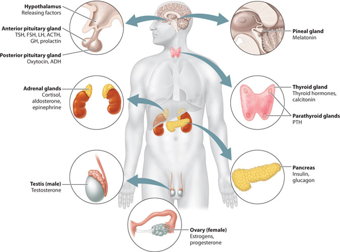
The pituitary gland communicates with many cells, tissues, and organs of the body. Some of these, like the thyroid and adrenal glands, secrete hormones in response to signals from the pituitary gland and therefore have exclusively endocrine functions. Others, like the lungs, kidneys, and digestive tract, harbor endocrine cells that secrete hormones and also have other physiological roles in the body. Consequently, the vertebrate endocrine system is not localized in one part of the body, but is present throughout.
38-13
38.3.1 The pituitary gland integrates diverse bodily functions by secreting hormones in response to signals from the hypothalamus.
The hypothalamus is the main route by which nervous system signals are transmitted to the vertebrate endocrine system. The function of the hypothalamus is to transmit these signals to the pituitary gland, the endocrine gland that acts as a control center for most other endocrine glands in the body.
The pituitary gland is divided into anterior and posterior regions (Fig. 38.11). This division is not arbitrary—the two regions have distinct functions, organizations, and embryonic origins. The anterior pituitary gland forms from epithelial cells that develop and push up from the roof of the mouth, whereas the posterior pituitary gland develops from neural tissue at the base of the brain. Both sets of ectodermal cells form pouches that develop into glands, which come to lie adjacent to each other. The anterior and posterior pituitary glands should therefore be thought of as two distinct glands, not one.
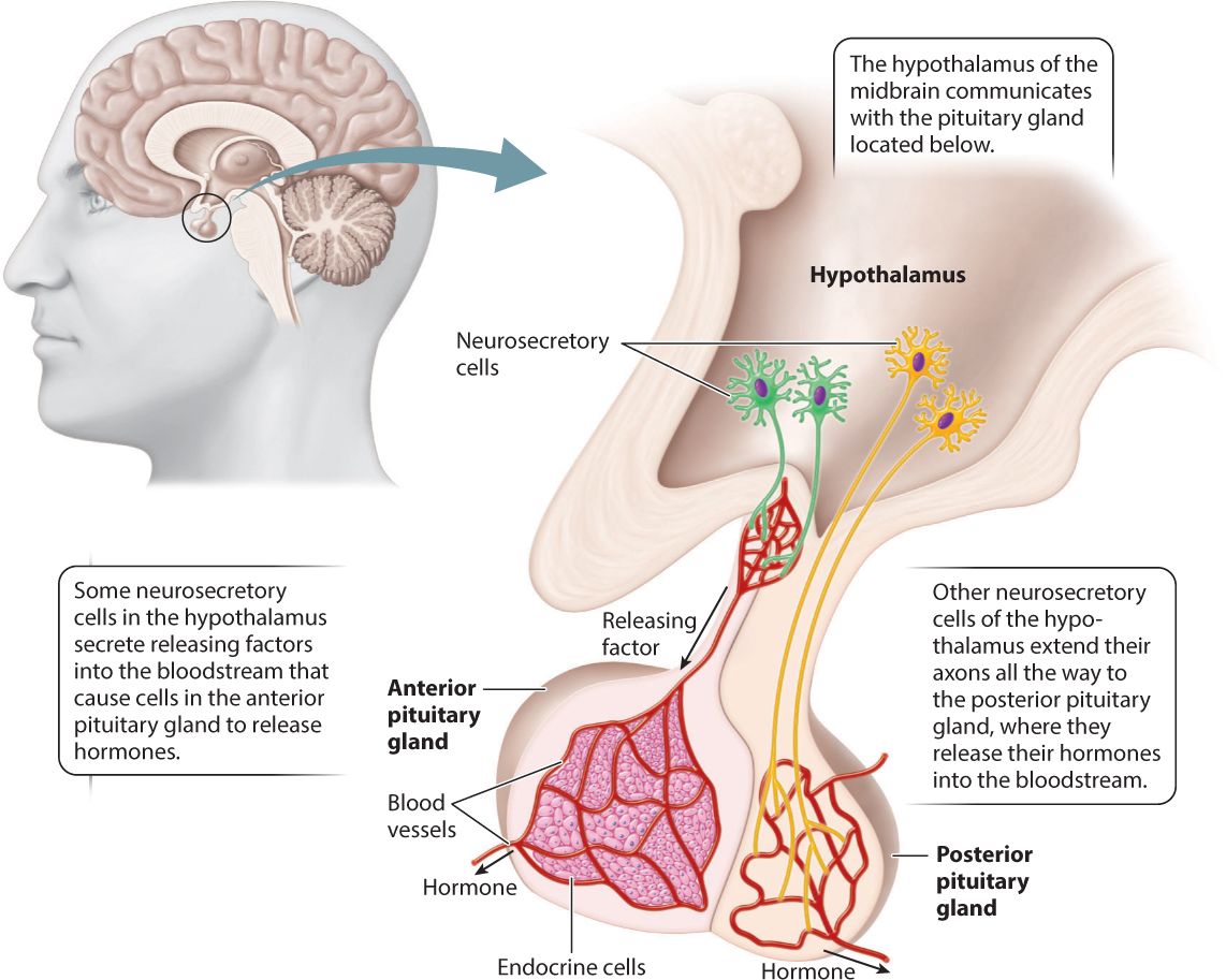
Because of these developmental differences, the anterior and posterior pituitary glands receive input from the hypothalamus in different ways. The hypothalamus contains neurosecretory cells. These cells are located in the hypothalamus, which is part of the brain, and therefore they are neurons. However, instead of secreting neurotransmitters that bind to another neuron or to muscle, these cells release hormones into the bloodstream. Some of these neurosecretory cells communicate with the anterior pituitary gland. In this case, they secrete hormones called releasing factors into small blood vessels that travel to and supply the anterior pituitary gland (Fig. 38.11). In response, cells of the anterior pituitary gland release hormones into the bloodstream, in which they circulate throughout the body and bind to receptors on target cells, tissues, and organs.
In contrast, communication between the hypothalamus and the posterior pituitary gland does not involve hypothalamic releasing factors. Instead, the posterior pituitary gland contains the axons of neurosecretory cells whose cell bodies are located in the hypothalamus. These axons release hormones directly into the bloodstream, by which they are transported to distant sites (Fig. 38.11). Consequently, the posterior pituitary is part of the nervous system itself.
In response to signals from the hypothalamus, distinct hormones are secreted by the anterior and posterior pituitary glands (Table 38.2). The anterior pituitary gland hormones include thyroid stimulating hormone (TSH), which acts on the thyroid gland, the gonadotropic hormones follicle-stimulating hormone (FSH) and luteinizing hormone (LH), which act on the female and male gonads (the ovaries and testes), and adrenocorticotropic hormone (ACTH), which acts on the adrenal glands (see Fig. 38.10). In each of these cases, the anterior pituitary hormone acts on an endocrine gland to cause release of other hormones. Hormones that control the release of other hormones are called tropic hormones. In response to TSH, the thyroid gland releases thyroid hormones that regulate the metabolic state of the body. In response to FSH and LH, the ovaries release estrogen and progesterone and the testes release testosterone. In response to ACTH, the adrenal glands release cortisol, which has diverse effects that include stimulating glucose release into the bloodstream, maintaining blood pressure, and suppressing the immune system.
38-14
The anterior pituitary gland also secretes growth hormone (GH), which acts generally on the muscles, bones, and other body tissues to stimulate their growth, and prolactin, which stimulates milk production in the breast of female mammals in response to an infant’s suckling at the mother’s nipple.
The posterior pituitary gland hormones include oxytocin and antidiuretic hormone (ADH; also called vasopressin). These are two evolutionarily related peptide hormones, each consisting of nine amino acids. As we discussed earlier, oxytocin plays several roles related to female reproduction: It causes uterine contraction during labor and stimulates the release of milk during breastfeeding. Antidiuretic hormone acts on the kidneys (Chapter 41), regulating the concentration of urine that an animal excretes, which is critical to maintaining water and solute balance in the body.
In addition to their roles in reproduction and kidney function, oxytocin and vasopressin are also released by cells in the brain and may play roles in social behaviors. For instance, oxytocin is important in regulating maternal behavior toward infants. Recent research has suggested that oxytocin may also have a role in behaviors such as trust as a prelude to mating in vertebrate animals. These functions reflect the long evolutionary history of oxytocin, as it also plays a role in the social behavior of insects.
Recent evidence suggests that antidiuretic hormone plays a parallel role in regulating male social behavior and parental behavior in mammals. This is not surprising because oxytocin and antidiuretic hormone have very similar chemical structures (see Fig. 38.7). The integration of reproductive function with parental and social behavior for the care of young is likely favored strongly by natural selection. Receptors for these hormones are expressed within the brain. Thus, in addition to acting on reproductive and renal organs of the body, oxytocin and antidiuretic hormone affect mammalian behavior by acting directly on the brain.
38.3.2 Many targets of pituitary hormones are endocrine tissues that also secrete hormones.
As we have seen, some of the hormones released by the anterior pituitary gland act on endocrine organs that themselves then release hormones. These tropic hormones are TSH, FSH and LH, and ACTH. Here, we look at their target organs in more detail.
TSH acts on the thyroid gland, which is located in the front of the neck (see Fig. 38.10), and leads to the release of two peptide hormones, thyroxine (T4) and triiodothyronine (T3). These hormones regulate cellular metabolism throughout the body. Overproduction of thyroid hormones (hyperthyroidism) or thyroid hormone deficiency (hypothyroidism) creates symptoms that reflect either an overly active metabolic state (increased appetite and weight loss) or one that is too low (fatigue and sluggishness). Both conditions are diagnosed by blood tests that monitor circulating levels of thyroid hormones, and both can be treated by medication.
Because thyroid hormones require iodine for their formation, individuals who do not acquire enough iodine from their diets produce too little thyroid hormone, resulting in metabolic problems. Additionally, iodine insufficiency stimulates increased production of TSH by the anterior pituitary gland because of the absence of negative feedback. Over time, the thyroid gland enlarges to form a goiter, which is observed as an enlargement of the throat, to compensate and produce more thyroid hormone. The introduction of iodized salt has eliminated goiter formation and hypothyroidism in many countries, but in many underdeveloped areas these remain significant public health problems.
The gonadotropic hormones are FSH and LH. They target the female and male gonads—the ovary and testis (see Fig. 38.10). In response to FSH and LH, the ovary and testis each secrete sex hormones that regulate their own development as well as the sexual differentiation and maturation of secondary sexual characteristics of the body. Female sex hormones include the steroids estrogen and progesterone. The principal male sex hormone is the androgen testosterone. These sex hormones are common to a vast majority of vertebrates, regulating sexual differentiation, gonadal maturation, and reproductive behavior. The role of these sex hormones in regulating reproductive function is discussed in greater detail in Chapter 42.
Testosterone is a naturally occurring anabolic steroid that stimulates the synthesis in the testes of proteins needed for sperm production and the development of male sexual features and body tissue growth, particularly in muscles. Anabolic steroids promote protein synthesis to build body tissues and anabolic metabolism to store energy within cells. A variety of synthetic anabolic steroids have been developed to treat muscle wasting due to loss of appetite and diseases such as cancer and AIDS. Anabolic steroids are used illegally by both male and female athletes to promote muscle strengthening, undermining fair competition. The risks to health of continued use of anabolic steroids are considerable: liver damage, heart disease, and high blood pressure. Tissue damage occurs because elevated circulating levels of testosterone produced by anabolic steroid use inhibits the normal synthesis of anterior pituitary tropic hormones.
38-15
ACTH released by the anterior pituitary gland is also a tropic hormone. It acts on the cortex (the outer portion) of the paired adrenal glands (see Fig. 38.10). The adrenal glands are located adjacent to the kidneys (“ad-” meaning “near” and “renal” referring to the kidneys). In humans, each small adrenal gland lies just above each kidney. During times of stress, such as starvation, fear, or intense physical exertion, ACTH stimulates adrenal cortex cells to secrete cortisol.
38.3.3 Other endocrine organs have diverse functions.
Although many endocrine organs receive signals from the hypothalamic–pituitary axis, some respond instead to internal physiological states of the body. One of these, adjacent to the thyroid gland, is the parathyroid gland (see Fig. 38.10), which secretes parathyroid hormone (PTH). Calcitonin, which is secreted by the thyroid gland, and PTH together regulate the actions of bone cells (osteoblasts and osteoclasts, Chapter 37) that control bone formation and bone removal and therefore the levels of calcium in the blood.
When circulating levels of calcium fall too low, the parathyroid gland triggers release of PTH, stimulating osteoclasts to reabsorb bone mineral, halting bone formation and raising blood calcium levels. When calcium levels are too high, the production of PTH is inhibited and calcitonin is released to shift bone metabolism toward net bone formation, building bone that stores calcium in the skeleton. Note that, as with blood glucose levels, calcium levels are maintained in a narrow range by negative feedback: The response to the hormone (increasing or decreasing levels of calcium in the blood) is the opposite of the stimulus (low or high levels of calcium in the blood) so that a stable set point is maintained.
Several organs of the digestive system, including the pancreas, stomach, and duodenum, produce hormones that regulate digestion, many of which are discussed in Chapter 40.
The pineal gland is located in the thalamic region of the brain (see Fig. 38.10). It responds to autonomic nervous system input by secreting melatonin, a hormone that helps control an animal’s state of wakefulness. When melatonin levels rise, animals sleep. In diurnal animals (those that are active during the day), increased levels occur at night; in nocturnal animals (those that are active during the night), increased levels occur during the day. The pineal gland is sensitive to changes in light and darkness. It releases melatonin in response to daily (and seasonal) light cycles, helping to maintain circadian rhythms of the body. Circadian rhythms are cycles of about 24 hours (or longer) in which an animal’s biochemical, physiological, and behavioral state shifts in response to changes in daily and seasonal environmental conditions. Exposure to sunlight inhibits the production of melatonin in diurnal animals, whereas darkness stimulates its release. To overcome jet lag, travelers are advised to seek sunlight and often to take melatonin as a medication before sleeping to help reset their 24-hour circadian rhythm. In other vertebrates, such as lampreys, fishes, and reptiles, the pineal gland functions as a “third eye,” sensing light through an opening in the skull. Light shining on the pineal gland regulates the animal’s general state of activity, influencing its behavioral response to changing light conditions.
In several animals, including ground squirrels, bats, bears, and rattlesnakes, the pineal gland regulates hibernation, controlling the animal’s metabolic state over longer time periods. In many animals, release of melatonin also regulates seasonal breeding cycles.
38.3.4 How does the endocrine system influence predators and prey?
Case 7 Predator–Prey: A Game of Life and Death
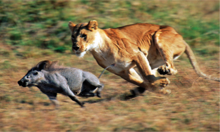
The endocrine system works closely with the nervous system to enable animals to respond to external cues in the environment. In Chapters 35 and 36, we discussed how prey animals, such as warthogs, use visual and olfactory cues processed by the nervous system to detect the distant movement of a predator, such as a lion, which in some instances occurs too late for a successful escape (Fig. 38.12). We saw how the sympathetic nervous system, in the fight-or-flight response, can trigger broad physiological changes, such as increased heart and breathing rates and changes in metabolism that ready the predator to attack or the prey to respond.
38-16
These actions are coordinated by the endocrine system. The sympathetic nervous system sends axons to the adrenal medulla, the inner part of the adrenal gland. In response to stimulation by the sympathetic nervous system, cells of the adrenal medulla secrete two hormones, epinephrine and norepinephrine (also known as adrenaline and noradrenaline). These are hormones released by the adrenal gland with targets throughout the body, leading to the physiological changes of the fight-or-flight response. Interestingly, epinephrine and norepinephrine also act as neurotransmitters in the brain. In fact, cells of the adrenal medulla are modified nerve cells that have lost their axons and dendrites.
The same set of responses occurs when, walking along a deserted sidewalk, we note a shadowy movement or hear a footstep behind us. The perceived threat sets in motion a coordinated physiological reaction, mediated by the combined action of the nervous and endocrine systems. These changes make us alert and ready for action to avoid or resist a perceived threat. They also inhibit digestive functions and eliminate a sense of appetite.
Earlier, we considered the importance of homeostasis in maintaining a variety of parameters, such as temperature and glucose levels, at a steady level. In this case, there is an adjustment of this set point, which is critical in enabling an organism to respond to external cues and adjust its physiological response appropriately. When the real or perceived threat is gone, the body is able to return to its prior state by reestablishing the earlier set point.