Chapter 2. Comparison of Cells: Prokaryotes and Eukaryotes
Objectives
By the end of the period, students will:
- practice using compound microscopes.
- be able to find and correctly focus on an object or a structure.
- be able to compare prokaryotic cells with eukaryotic cells.
- be able to recognize and compare animal, plant, and Protista cells.
- be able to recognize the stages of mitosis in cell division.
Useful Website
Using Microscopes to Study Cells
This lab is a continuation of the last week’s lab and is designed to give students more practice using microscopes and the opportunity to look at a wider variety of cells.
Your TA will start with a quick reminder of how to use the microscopes correctly!
All living things, including humans, are composed of cells, but cells differ enormously in shape, size, and capabilities. For example, in humans and other animals, there are cells that store fat (fat cells), cells that transmit electrical impulses (nerve cells), cells that secrete digestive enzymes or chemical messengers (epithelial cells), cells specialized for absorption of light, cells that secrete the mineral constituents of bone, cells that protect against infection, cells with hairs, cells that move by swimming, and cells that move by crawling. The largest (or at least longest) cell is a neuron or nerve cell found in a giraffe. It may be as many as four meters long!
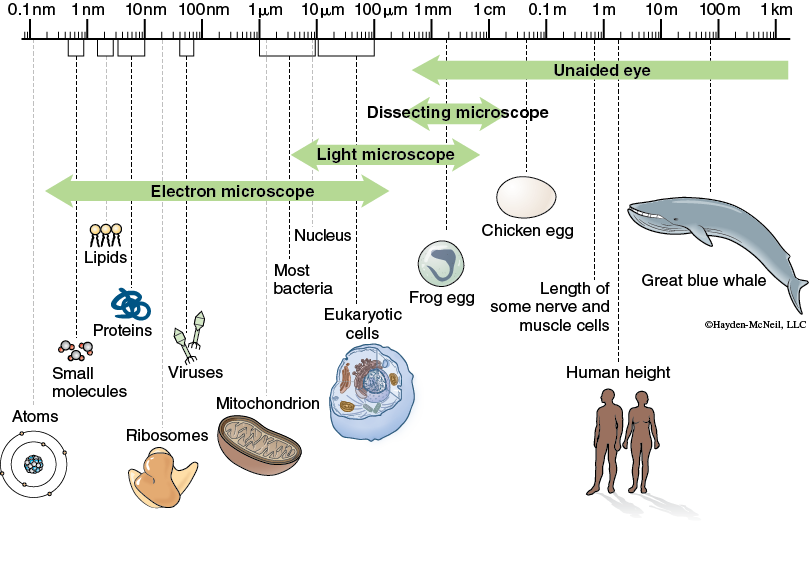
Cells, no matter what their function, can be grouped into two major categories. The cells of bacteria are prokaryotic. Prokaryotic cells are very small (0.2–5 microns; note that there are 1000 microns in a millimeter) and lack a true nucleus or other membrane-bound organelles. Eukaryotic cells tend to be larger (10–100 microns long) and have a true nucleus and other membrane-bound organelles. The largest eukaryotic cells, such as an amoeba, might be almost visible without a microscope. All organisms except bacteria are composed of eukaryotic cells. The nucleus contains the DNA organized into chromosomes. Figure 4.1 shows the range of sizes that are relevant to people and the ranges of sizes for which microscopes are helpful.
Part 1. Comparing Prokaryotic and Eukaryotic Cells
We will start by looking at prokaryotic cells. There will be a demonstration set up of bacterial cells at 100× magnification. Recall that at 100× we need to use oil between the lens and the slide. In order to see how much more you can see at 100×, we will also have a demo set up at 40×.
We will also look at an example of cyanobacteria, a type of prokaryote that is common in freshwater ponds. For each of these, note that there are no organelles that are visible and no nucleus.
Making a Wet Mount
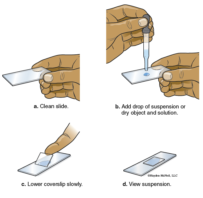
Many small objects are best viewed by immersing the specimen in water in a preparation called a wet mount, and this is what we will use to look at the cyanobacteria. The materials that you need will be on each table.
- Obtain a clean microscope slide and coverslip. If they are not clean, use a paper towel (not lens paper) to clean them. (Lens paper is too expensive to be used for this menial task—reserve it for the lenses.) While holding the slide level, put a drop of water on the center of the slide.
- Take a drop of water from the “pond water” provided in class.
- Add a coverslip as indicated in Figure 4.2. If water does not completely fill the area under the slide, place a small drop of water somewhere on the slide so that it just touches the edge of the coverslip and it will be drawn under the coverslip. Now, look at what you see!
Part 2. Comparing Various Eukaryotic Cells (Plants, Animals, Protista, and Fungi)
In BISC 162 and BISC 163, we will do a much more thorough comparison of these groups of organisms. For this semester, we want to use examples of each of these groups to reinforce the notion that everything is made of cells and that eukaryotic cells are very different from prokaryotic cells. We also want you to know some of the key differences between the cells of each of these groups.
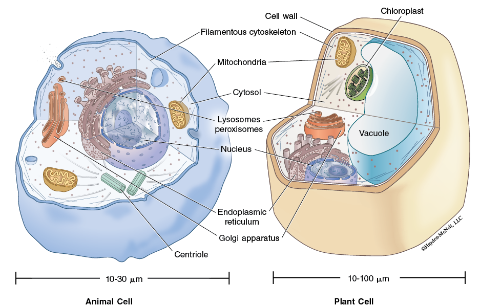
Plant cells have a cell wall in addition to a cell membrane. This makes cells fairly “boxy” on appearance. They also tend to have a large central fluid filled vacuole. Chloroplasts, the green organelles of photosynthesis, are often visible, as is the nucleus.
Animal cells also have a nucleus (they are eukaryotes, after all) but lack the cell wall, vacuole, and chloroplasts. Animal cells take a wide variety of shapes depending on their function. Figure 4.3 shows a generalized animal cell and plants cell.
There is no such thing as a typical protist, so we will have a couple of examples available for you to look at. Some have chloroplasts (and photosynthesize), some do not.
Fungi tend to grow in long filaments called hyphae. They also have cell walls, but these walls are made of chitin rather than cellulose, as is the case with plant cell walls.
We want to you to look at examples of each of these and make sketches of each. How are they similar? How are they diferent?
Part 3. Mitosis and the Cell Cycle
Cells normally undergo a complex series of steps that results in cells growing and dividing. The control of this process is complex and details about the control are still being discovered. Cells that grow uncontrollably are cancer cells. In lecture, we will cover this process in some detail. In lab, we want you to be able to find and recognize the different stages of mitosis.
Mitosis is when the replicated chromosomes separate and cytokinesis divides the cytoplasm to the two daughter cells. Mitosis is divided into four stages:
Interphase encompasses the G1 (growth 1) Phase, S (DNA Synthesis Phase 4) and G2 (growth 2) Phase.
Prophase: During prophase, the chromatin condenses and become visible as chromosomes.
Metaphase: Chromosomes align along the equatorial plane.
Anaphase: The paired chromosomes separate at the kinetochores and move to opposite poles.
Telophase: Chromatids arrive at the opposite poles and new nuclear membranes form.
Cytokinesis: Division of the cytoplasm occurs when a protein pinches the cell into two daughter cells.
Instructions: Get a slide of the onion root tip. On the slides, find examples of each of the phases. When you have found an example, raise your hand for the TA to confirm that you have found it. Be prepared to explain why you think that cell is in that stage. On Hand-In 4, make a sketch of what each stage looks like.
CHECKLIST FOR THE END OF LAB
When you are finished, each lab must be clean and ready for the next group of students.
Notes to Instructors and Materials for Lab
TAs will set up a demo of a bacterial slide. It will be helpful to have two scopes set up— one at 100× with oil and the other at 40×.
We will have Oscillatoria as an example of cyanobacteria.
Our plant cells will be Elodea, a common aquatic plant.
Animal cells will be epithelial cells.
Protists will include a green algae (Chlamydomonas) and Paramecium.
We have prepared slides of a variety of fungus.
We will use onion root tip slides for mitosis.