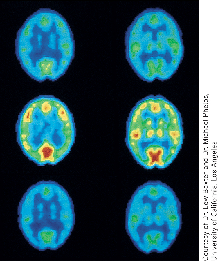
Brain Activity During the Extremes of Bipolar Disorder These PET scans record the brain activity of an individual with bipolar disorder as he cycled rapidly from depression to mania and back to depression over a 10-day period. In the top and bottom PET scans, the blue and green colors clearly show the sharp reduction in overall brain activity that coincided with the episodes of depression. In the center PET scans, the bright red, orange, and yellow colors indicate high levels of activity in diverse brain regions during the intervening episodes of mania.
Source: Lewis Baxter and Michael E. Phelps, UCLA School of Medicine.
Courtesy of Dr. Lew Baxter and Dr. Michael Phelps, University of California, Los Angeles
Courtesy of Dr. Lew Baxter and Dr. Michael Phelps, University of California, Los Angeles