2.5 Specialization in the Cerebral Hemispheres
KEY THEME
Although they have many functions in common, the two hemispheres of the cerebral cortex are specialized for different tasks.
KEY QUESTIONS
How did Broca, Wernicke, and Sperry contribute to our knowledge of the brain?
Why would the corpus callosum be surgically severed, and what effects would that produce?
How do the functions of the right and left cerebral hemispheres differ?
!launch!
If you hold a human brain in your hand, the two cerebral hemispheres would appear to be symmetrical. Although the left and right hemispheres are very similar in appearance, they are not identical. Anatomically, one hemisphere may be slightly larger than the other. There are also subtle differences in the sizes of particular structures, in the distribution of gray matter and white matter, and in the patterns of folds, bulges, and grooves that make up the surface of the cerebral cortex (Ocklenburg & Güntürkün, 2012).
What about differences in the functions of the two hemispheres? In many cases, the functioning of the left and right hemispheres is symmetrical, meaning that the same functions are located in roughly the same places on each hemisphere. Examples of such functional symmetry include the primary motor cortex and the somatosensory cortex, which we discussed in the previous section. With regard to other important processes, however, the left and right cerebral hemispheres do differ—each cerebral hemisphere is specialized for particular abilities.
Here’s a rough analogy. Imagine two computers that are linked through a network. One computer is optimized for handling word processing, the other for handling graphic design. Although specialized for different functions, the two computers actively share information and can communicate with each other across the network. In this analogy, the two computers correspond to the left and right cerebral hemispheres, and the network that links them is the corpus callosum.
CRITICAL THINKING
“His” and “Her” Brains?
Do men and women have fundamentally different brains, “hardwired” by hormones and genes to produce what some popular authors claim are “separate realities” (Brizendine, 2006, 2010)? According to this view, there are innate, unchangeable differences between the brains of males and females, and these differences cause gender differences in behaviors, attitudes, personality traits, and skills (Fine, 2012). But what are these differences? Do they cause men and women to “think, feel, and behave differently,” as some headlines and popular books claim? Let’s look at the scientific research.
MYTH !lhtriangle! SCIENCE
Is it true that because their brains are wired differently, men and women think, feel, and behave differently?
Researchers have found numerous sex differences in brain structure and function (Cahill, 2012, 2014; McCarthy & others, 2012). For example, men’s brains tend to be larger than women’s brains, mainly because their skulls also tend to be larger. And, women tend to have a higher proportion of gray matter to white matter in their brains than men. Gray matter and white matter are also distributed differently. In females, the distribution of gray and white matter is very similar in the left and right hemispheres. But males have a higher percentage of gray matter in their left hemisphere than in their right hemisphere (Halpern & others, 2007).
In general, the male brain is more asymmetrical and functions are more lateralized than in the female brain (Tomasi & Volkow, 2012a, b, 2011; Tiedt & others, 2013). For example, some studies have found that women tend to rely on brain structures in both hemispheres for certain language tasks, while men are more reliant on one brain hemisphere, usually the left, for language tasks (Cahill, 2006). Similarly, another study found that males rely more on focused activation of specific brain regions in the right hemisphere for visual and spatial tasks (Clements, 2006).
But what do such differences mean? Some researchers speculate that such differences may explain females’ stronger language skills and male’s stronger spatial skills (Tomasi & Volkow, 2012). But even neuroscientists are not sure, pointing to other studies that show no difference between the sexes, or that show females being more lateralized than males for some tasks (Bourne & Maxwell, 2010; Fine, 2012b).
Thinking Critically About Brain Differences
How should you interpret media sound-bites about profound sex differences in the brain? Or claims that certain attitudes or behaviors are “hard-wired in the brain”?
First, it’s important to think critically about media claims. Unlike reporters, scientists are usually careful to qualify their conclusions and describe the limitations of their research. These limitations are rarely mentioned in media reports. For example, brain studies are typically based on small groups of participants, who may or may not be representative of the wider population of men and women. And, while findings of sex differences in brain structure or function tend to be widely reported in the media, findings of no difference go unreported. So, often, does the failure of studies to be replicated by other researchers (Eliot, 2011; Fine, 2013a, 2013b). In other words, the findings of individual research studies are rarely as earth-shattering as reported.
Second, most sex differences amount to minor variations in a particular brain region or to statistical differences in the average level of activation of particular brain regions. Brain structures and functioning are essentially the same in men and women (Jordan-Young & Rumiati, 2011). As you’ll see in Chapter 10, on sex and gender, when it comes to personality traits, abilities, and attitudes, men and women are much more similar than they are different.
The hardwiring paradigm erases the effect of the social world in producing sex/gender differences, so that sex/gender hierarchies appear natural. Neuroscientific explanations of sex/gender differences have added a new allure to an old fashioned sexism.
—Rebecca Jordan-Young & Raffaella I. Rumiati (2011)
Finally, it’s important to remember that even differences that are biological in origin are not necessarily fixed, permanent, or inevitable (Fine, 2013a, b). Brain development and function are affected by biological influences, such as exposure to the sex hormones produced both before birth and throughout your life (Lombardo & others, 2012). However, these biological factors themselves are strongly influenced by environmental factors, ranging from the food we eat to the stressful circumstances we experience. As we’ve emphasized throughout this chapter, both brain function and structure are highly responsive to environmental influences (Fine & others, 2013). Thus, sex differences in structures or function might well be the result of the different life experiences of men and women, rather than the cause (Fine & others, 2013; Kaiser, 2012).
CRITICAL THINKING QUESTIONS
Why are sweeping claims about fundamental sex differences in the human brain misleading?
What is wrong with the statement that certain behaviors or personality traits are “hard-wired” in the male or female brain?
How might claims that sex differences are “hard-wired” or innate affect attitudes or behavior?
Why is the notion that sex differences might be due to brain differences so appealing to many people?
As you’ll see in this section, the first discoveries about the differing abilities of the two brain hemispheres were made more than a hundred years ago by two important pioneers in brain research, Pierre Paul Broca and Karl Wernicke.
Language and the Left Hemisphere: THE EARLY WORK OF BROCA AND WERNICKE
By the end of the 1700s it had already been well established that injury to one side of the brain could produce muscle paralysis or loss of sensation on the opposite side of the body. By the early 1800s, animal experiments had shown that specific functions would be lost if particular brain areas were destroyed. And, as discussed in the Science Versus Pseudoscience box on page 63, phrenology triggered scientific debates about cortical localization, or localization of function—the idea that particular brain areas are associated with specific functions.
cortical localization
The notion that different functions are located or localized in different areas of the brain; also called localization of function.
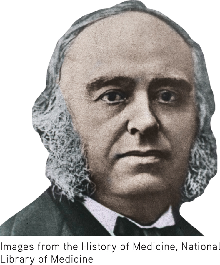
In the 1860s, more compelling evidence for cortical localization was presented by a French surgeon and neuroanatomist named Pierre Paul Broca. Broca treated a series of patients who had great difficulty speaking but could comprehend written or spoken language. Subsequent autopsies of these patients revealed a consistent finding—brain damage to an area on the lower left frontal lobe. Today, this area on the left hemisphere is referred to as Broca’s area, and it is known to play a crucial role in speech production (FIGURE 2.21).
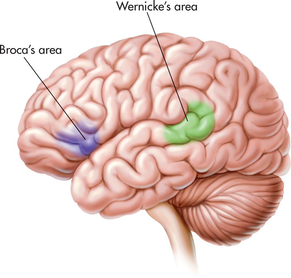

About a decade after Broca’s discovery, a young German neurologist named Karl Wernicke discovered another area in the left hemisphere that, when damaged, produced a different type of language disturbance. Unlike Broca’s patients, Wernicke’s patients had great difficulty understanding spoken or written communications. They could speak quickly and easily, but their speech sometimes made no sense. They sometimes used meaningless words or even nonsense syllables, though their sentences seemed to be grammatical. In response to the question “How are you feeling?” a patient might say something like, “Don’t glow glover. Yes, uh, ummm, bick, bo chipickers the dallydoe mick more work mittle.” Autopsies of these patients’ brains revealed consistent damage to an area on the left temporal lobe that today is called Wernicke’s area (see FIGURE 2.21).
The discoveries of Broca and Wernicke provided the first compelling clinical evidence that language and speech functions are performed primarily by the left cerebral hemisphere. If similar brain damage occurs in the exact same locations on the right hemisphere, these severe disruptions in language and speech are usually not seen.
The notion that one hemisphere exerts more control over or is more involved in the processing of a particular psychological function is termed lateralization of function. Speech and language functions are lateralized on the left hemisphere. Generally, the left hemisphere exerts greater control over speech and language abilities in virtually all right-handed and the majority of left-handed people.
lateralization of function
The notion that specific psychological or cognitive functions are processed primarily on one side of the brain.
The language disruptions demonstrated by Broca’s and Wernicke’s patients represent different types of aphasia. Aphasia refers to the partial or complete inability to articulate ideas or understand spoken or written language because of brain injury or damage. There are many different types of aphasia.
aphasia
(uh-FAYZH-yuh) The partial or complete inability to articulate ideas or understand spoken or written language because of brain injury or damage.
People with Broca’s aphasia find it difficult or impossible to produce speech, which is why it is often referred to as expressive aphasia. Despite their impairments in speaking, their comprehension of verbal or written words is relatively unaffected.
People with Wernicke’s aphasia have great difficulty comprehending written or spoken communication, which is why it is often referred to as receptive aphasia. Although they can speak, they often have trouble finding the correct words.

At the beginning of this chapter, we described the symptoms experienced by our friend Asha in the weeks before and the months following her stroke. Asha, who is right-handed, experienced the stroke in her left hemisphere. About three days after her stroke, an MRI brain scan showed where the damage had occurred: the left temporal lobe. Asha experienced many symptoms of Wernicke’s aphasia. Talking was difficult, not because Asha couldn’t speak, but because she had to stop frequently to search for the right words. Asha was unable to name even simple objects, like the cup on her hospital dinner tray or her doctor’s necktie. She recognized the objects but was unable to say what they were. She had great difficulty following a normal conversation and understanding speech, both in English and in her native language, Tulu.
Asha also discovered that she had lost the ability to read. She could see the words on the page, but they seemed to have no meaning. Her husband, Paul, brought some of their Christmas cards to the hospital. Asha recalls, “When I realized I couldn’t read the Christmas cards, I thought my life was over. I just lost it. I remember crying and telling the nurse, ‘I have a doctorate and I can’t read, write, or talk!’”
When we visited Asha in the hospital, we brought her a Christmas present: a portable music player with headphones and some albums of relaxing instrumental music. Little did we realize how helpful the music would be for her. One album was a recording of Native American flute music called Sky of Dreams. The music was beautiful and rather unusual, with intricate melodies and unexpected, complex harmonies. Although it was very difficult for Asha to follow normal speech, listening to Sky of Dreams was an entirely different experience. As Asha explained:
I tried cranking up the music very high and it soothed me. I could sleep. At the time, the flute music seemed to be just perfectly timed with the way my brain was working. It was tuning out all the other noises so I could focus on just one thing and sleep. So I would play the music over and over again at a very high level. I did that for a long time because my mind was so active and jumbled that I couldn’t think.
Asha’s language functions were severely disrupted, yet she was able to listen to and appreciate instrumental music—even very complex music. Why? At the end of the next section, we’ll offer a possible explanation for what seems to have been a disparity in Asha’s cognitive abilities following her stroke.
Cutting the Corpus Callosum: THE SPLIT BRAIN
Since the discoveries by Broca and Wernicke, the most dramatic evidence illustrating the independent functions of the two cerebral hemispheres has come from a surgical procedure called the split-brain operation. This operation is used to stop or reduce recurring seizures in severe cases of epilepsy that can’t be treated in any other fashion. The procedure involves surgically cutting the corpus callosum, the thick band of axons that connects the two hemispheres.
split-brain operation
A surgical procedure that involves cutting the corpus callosum.
What was the logic behind cutting the corpus callosum? An epileptic seizure typically occurs when neurons begin firing in a disorganized fashion in one region of the brain. The disorganized neuronal firing quickly spreads from one hemisphere to the other via the corpus callosum. If the corpus callosum is cut, seizures should be contained in just one hemisphere, reducing their severity or eliminating them altogether. This is exactly what happened when the split-brain operation was first tried in this country in the 1940s (Springer & Deutsch, 1998).
Surprisingly, cutting the corpus callosum initially seemed to produce no noticeable effect on the patients, other than reducing their epileptic seizures. Their ability to engage in routine conversations and tasks seemed to be unaffected. On the basis of these early observations, some brain researchers speculated that the corpus callosum served no function whatsoever. One famous psychologist, Karl Lashley, joked that the primary function of the corpus callosum seemed to be to keep the two hemispheres from sagging (Hoptman & Davidson, 1994).
In the 1960s, however, psychologist and neuroscientist Roger Sperry and his colleagues began unraveling the puzzle of the left and right hemispheres. Sperry and his colleagues used the apparatus shown in FIGURE 2.22 to test the abilities of split-brain patients. They would direct a split-brain subject to focus on a point in the middle of a screen, while briefly flashing a word or picture to the left or right of the midpoint.
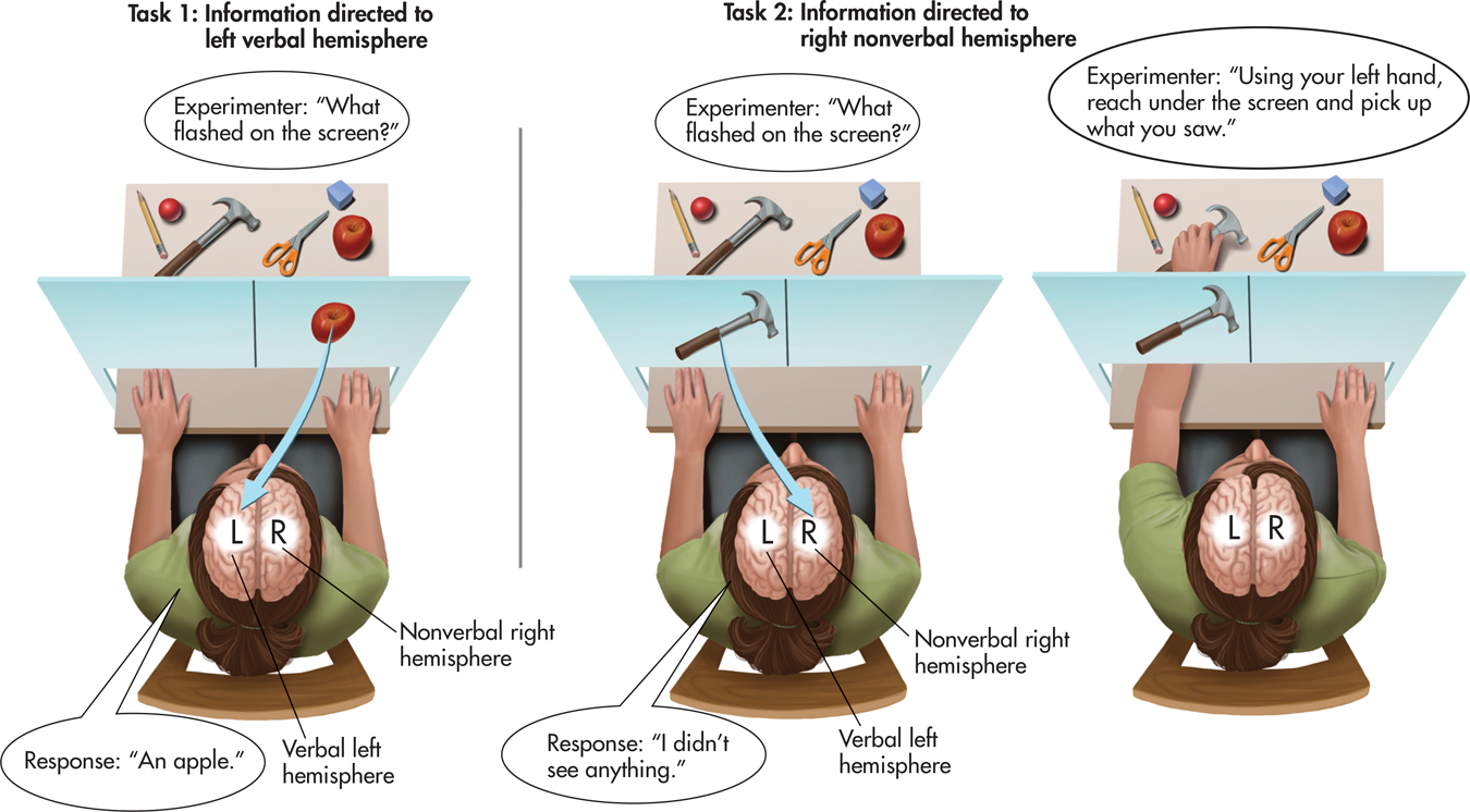
In this procedure, visual information to the right of the midpoint is projected to the person’s left hemisphere, and visual information to the left of the midpoint is projected to the right hemisphere. Behind the screen several objects were hidden from the split-brain subject. The subject could reach under a partition below the screen to pick up the concealed objects but could not see them (Sperry, 1982).
In a typical experiment, Sperry projected the image of an object concealed behind the screen, such as a hammer, to the left of the midpoint. This is shown in Task 2, FIGURE 2.22. Thus, the image of the hammer was sent to the right, nonverbal hemisphere. If a split-brain subject was asked to verbally identify the image flashed on the screen, she could not do so and often denied that anything had appeared on the screen. Why? Because her verbal left hemisphere had no way of knowing the information that had been sent to her right hemisphere. However, if a split-brain subject was asked to use her left hand to reach under the partition for the object that had been displayed, she would correctly pick up the hammer. This was because her left hand was controlled by the same right hemisphere that saw the image of the hammer.
Sperry’s experiments reconfirmed the specialized language abilities of the left hemisphere that Broca and Wernicke had discovered more than a hundred years earlier. But notice, even though the split-brain subject’s right hemisphere could not express itself verbally, it still processed information and expressed itself nonverbally: The subject was able to pick up the correct object.
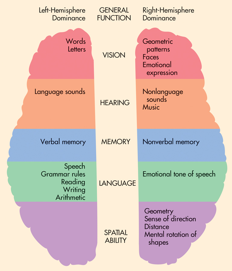
Over the last four decades, researchers have gained numerous insights about the brain’s lateralization of functions by studying split-brain patients, using brain-imaging techniques with normal subjects, and employing other techniques (Gazzaniga, 2005; Hugdahl & Westerhausen, 2010). On the basis of this evidence, researchers have concluded that—in most people—the left hemisphere is superior in language abilities, speech, reading, and writing.
In contrast, the right hemisphere is more involved in nonverbal emotional expression and visual-spatial tasks (Corballis, 2010). Deciphering complex visual cues, such as completing a puzzle or manipulating blocks to match a particular design, also relies on right-hemisphere processing (Gazzaniga, 1995, 2005). And the right hemisphere excels in recognizing faces and emotional facial cues, reading maps, copying designs, and drawing. Finally, the right hemisphere shows a higher degree of specialization for musical appreciation or responsiveness—but not necessarily for musical ability, which involves the use of the left hemisphere as well (Springer & Deutsch, 2001).
Think Like a SCIENTIST
Can you be classified as right-brained or left-brained? Go to LaunchPad: Resources to Think Like a Scientist about The Right Brain Versus the Left Brain.

FIGURE 2.23 summarizes the research findings for the different specialized abilities of the two hemispheres for right-handed people. As you look at the figure, it’s important to keep two points in mind. First, the differences between the left and right hemispheres are almost always relative differences, not absolute differences. In other words, both hemispheres of your brain are activated to some extent as you perform virtually any task (Toga & Thompson, 2003). In the normal brain, the left and right hemispheres function in an integrated fashion, constantly exchanging information (Allen & others, 2007; Banich, 1998). Thus, FIGURE 2.23 indicates the hemisphere that typically displays greater activation or exerts greater control over a particular function. Misconceptions about the roles played by the left and right hemispheres are common in the popular media. The Science Versus Pseudoscience box, “Brain Myths,” explores some of the most common misperceptions about the brain. Second, many functions of the cerebral hemispheres, such as those involving the primary sensory and motor areas, are symmetrical. They are located in the same place and are performed in the same way on both the left and the right hemispheres.
SCIENCE VERSUS PSEUDOSCIENCE
Brain Myths
Is it true that some people are “right-brained” and other people “left-brained”?
To investigate this question, researchers compared more than a thousand fMRI scans taken while participants rested. There was no evidence that some participants differed in their average levels of left and right hemisphere activation, as you would expect if some people were “left-brained” and others “right-brained” (Nielsen & others, 2013).
It certainly seems as if some people are more logical, analytical, or detail-oriented than others, especially in the way that they make decisions or tackle problems. But remember that the left and right hemispheres are highly interconnected in the normal, intact human brain. Unless the corpus callosum has been surgically sliced, all humans rely on the smooth, integrated functioning of both their left and right hemispheres to speak, learn, and generally navigate everyday life. In fact, the more complex the task, the greater the likelihood that both hemispheres will be involved in performing it (Allen & others, 2007; Yoshizaki & others, 2007).
What about left-handed people? Is it true that they are right-hemisphere-dominant?
Only about 10 to 13 percent of the population identify themselves as left-handed (Basso, 2007). Unlike right-handed people, who tend to use their right hands for virtually all tasks requiring dexterity, most left-handers actually show a pattern of “mixed” handedness. For example, your author Don uses his left hand to write and hold a fork but his right hand to swing a tennis racquet. Strong left-handedness is extremely rare (Wolman, 2005).
It’s a myth that left-handers have a fundamentally different brain organization from right-handers. About 75 percent of left-handers are left-hemisphere-dominant for language, just like right-handers. The remaining 25 percent are either right-hemisphere-dominant for language or bilateral, using both hemispheres for speech and language functions. Just for the record, about 5 percent of right-handed people are also either right-hemisphere or bilaterally specialized for language (Knecht & others, 2000; Ocklenburg & Güntürkün, 2012).
Is the right brain responsible for creativity and intuition? Can you train your right brain?
MYTH !lhtriangle! SCIENCE
Is it true that the right brain is creative and intuitive, and the left brain is analytic and logical, but that left-brained people can educate their right brain?
Although the right hemisphere is specialized for holistic processing, there is no evidence that the right hemisphere is any more “intuitive” or “creative” than the left hemisphere (Gazzaniga, 2005). In fact, a recent study found that a task requiring a creative solution involved greater left hemisphere activation than a task that required a noncreative solution (Aziz-Zadeh & others, 2013). There is also no evidence that any teacher, however skilled, could somehow selectively “educate” one side of your brain in isolation from the other (Goswami, 2006). While it is true that each hemisphere is specialized for different abilities, you rely on the integrated functioning of both hemispheres to accomplish most tasks. This is especially true for such cognitively demanding tasks as artistic creativity, musical performance, or finding innovative solutions to complex problems.
Given the basic findings on the laterality of different functions in the two hemispheres, can you speculate about why Asha was unable to read or follow a simple conversation but could easily concentrate on a complex piece of music? Why were her language abilities so disrupted, while her ability to focus on and appreciate music remained intact after her stroke?
A plausible explanation has to do with the location of the stroke’s damage on Asha’s left temporal lobe. Because language functions are usually localized on the left hemisphere, the stroke produced serious disruptions in Asha’s language abilities. However, her right cerebral hemisphere sustained no detectable damage. Because one of the right hemisphere’s abilities is the appreciation of musical sounds, Asha retained the ability to concentrate on and appreciate music.
Question 2.13
0gamQ66gkYANNs9yREumEjiMGq62smBY0dhnO7ExNTntVGu4FjAnHHiTIjADVc3jrB/T2spPBlwCKmPar91Jrfyqf4+duT2YAhMBuzmgZM/fC9qNNqTBjYD6I9ThFCkko13M4CHgWGRyVdviib0oWkQ4WkfIXRCZ/andptuW9hC0D7vOK44An1ujXcLduginVN4762PeduOY05qbqTYqg7QN7/cg7CApk47ZGukDUbKXHST6OOl1pflvFlrQiXS16+bVPqaBHSRoeqkGRla1RA==CONCEPT REVIEW 2.3
The Brain
Fill in the blanks in the following statements.
Question 2.14
1. The three major regions of the brain are the ____________ , ____________ , and the ____________ .
Question 2.15
2. Coordinated muscle movements and balance are regulated by the ____________
Question 2.16
3. Auditory information is processed in the ____________ lobe.
Question 2.17
4. The signals for voluntary movements originate in the ____________ , which is located on the ____________ lobe.
Question 2.18
5. The ____________ plays a critical role in the formation of new memories.
Question 2.19
6. The hypothalamus, hippocampus, amygdala, and parts of the thalamus and frontal cortex make up the ____________ .
Question 2.20
7. Damage to ____________ on the brain’s left hemisphere disrupts the ability to speak but not the ability to understand spoken or written communication.
Question 2.21
8. The ____________ is surgically cut in the split-brain operation.
Question 2.22
9. Our daily sleep–
Test your understanding of The Brain with with
 .
.
Closing Thoughts
In our exploration of neuroscience and behavior, we’ve traveled from the activities of individual neurons to the complex interaction of the billions of neurons that make up the human nervous system, most notably the brain. In the course of those travels, we presented four themes that are crucial to a scientific understanding of brain function: localization, lateralization, integration, and plasticity.

More than just a historical scientific oddity, phrenology’s incorrect interpretation of bumps on the skull helped focus scientific debate on the notion of localization—the idea that different functions are localized in different brain areas. Although rejected in the early 1800s when Franz Gall was in his heyday, localization of brain functioning is well established today. The early clinical evidence provided by Broca and Wernicke, and the later split-brain evidence provided by Sperry and his colleagues, confirmed the idea of lateralization—that some functions are performed primarily by one cerebral hemisphere.
The ideas of localization and lateralization are complemented by another theme evident in this chapter—integration. Although the nervous system is highly specialized, even simple behaviors involve the highly integrated interaction of trillions of synapses. Your ability to process new information and experiences, your memories of previous experiences, your sense of who you are and what you know, your actions and reactions—all depend upon the harmony of the nervous system.
The story of Asha’s stroke illustrated what can happen when that harmony is disrupted. Asha survived her stroke, but many people who suffer strokes do not. Of those who do survive a stroke, about one-third are left with severe impairments in their ability to function.
What happened to Asha? Fortunately, her story has a happy ending. Asha was luckier than many stroke victims—she was young, strong, and otherwise healthy. Asha’s recovery was also aided by her high level of motivation, willingness to work hard, and sheer will to recover. After being discharged from the hospital, Asha began months of intensive speech therapy. Her speech therapist assigned a great deal of homework that consisted of repeatedly pairing pictures with words, objects with words, and words with objects. Asha was literally rewiring her brain by relearning the correct associations between words and their meanings.
Asha set a very high goal for herself: to return to teaching at the university the following fall semester. With the help of her husband, Paul, and her mother, Nalini, who traveled from India to help coach her back to full recovery, Asha made progressive and significant gains. With remarkable determination, Asha reached the goal she had set for herself. Eight months after her stroke, she returned to the classroom and her research lab.
Today, more than five years after her stroke, the average person would never know that Asha had sustained significant brain damage. Other than an occasional tendency to “block” on familiar words—especially when she’s very tired—Asha seems to have made a complete recovery.
Thus, Asha’s story illustrates a final theme—the brain’s remarkable plasticity. Next, we take a closer look at how the brain responds to different types of environments. You will also learn how you can use research to enhance your own dendritic potential!