Gel electrophoresis
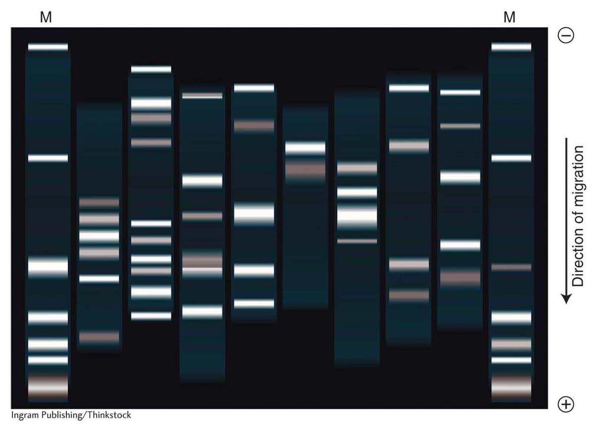
Mixtures of different-size DNA fragments have been separated electrophoretically on an agarose gel. The samples are eight recombinant vectors treated with EcoRI. The mixtures are applied to wells near the top of the gel, and fragments move from the negative to the positive end under the influence of an electrical field to different positions dependent on size. The DNA bands have been visualized by staining with ethidium bromide and photographing under ultraviolet light. (The letter M represents lanes containing standard fragments acting as markers for estimating DNA length.)
[Ingram Publishing/Thinkstock]