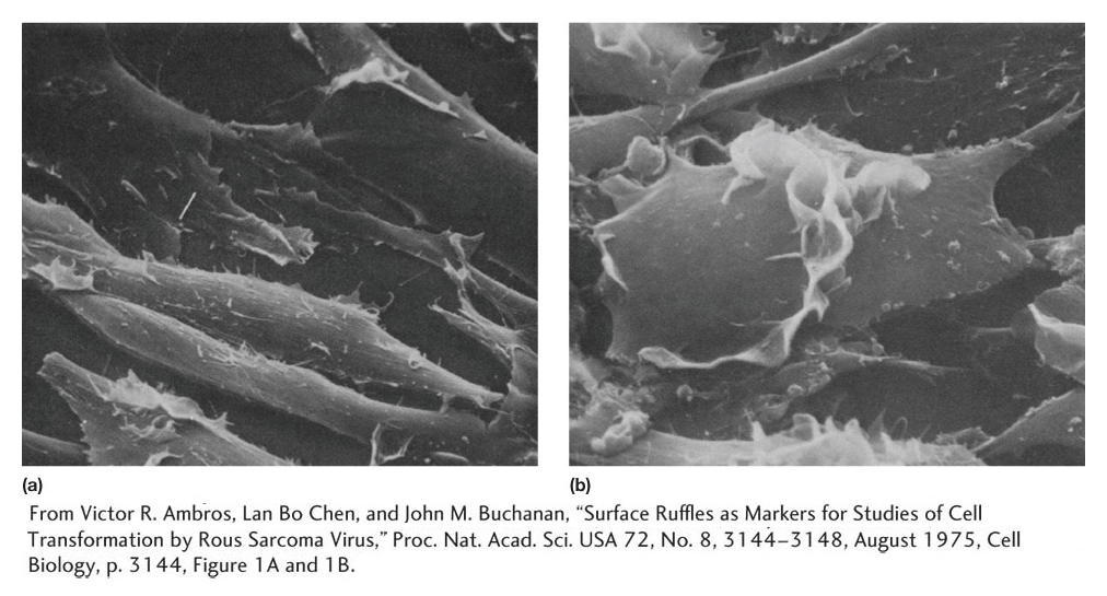
Scanning electron micrographs of (a) normal cells and (b) cells transformed by Rous sarcoma virus, which infects cells with the src oncogene. (a) A normal cell line called 3T3. Note the organized monolayer structure of the cells. (b) A transformed derivative of 3T3. Note how the cells are rounder and piled up on one another.
[From Victor R. Ambros, Lan Bo Chen, and John M. Buchanan, “Surface Ruffles as Markers for Studies of Cell Transformation by Rous Sarcoma Virus,” Proc. Nat. Acad. Sci. USA 72, No. 8, 3144-