Donor DNA is transferred as a single strand
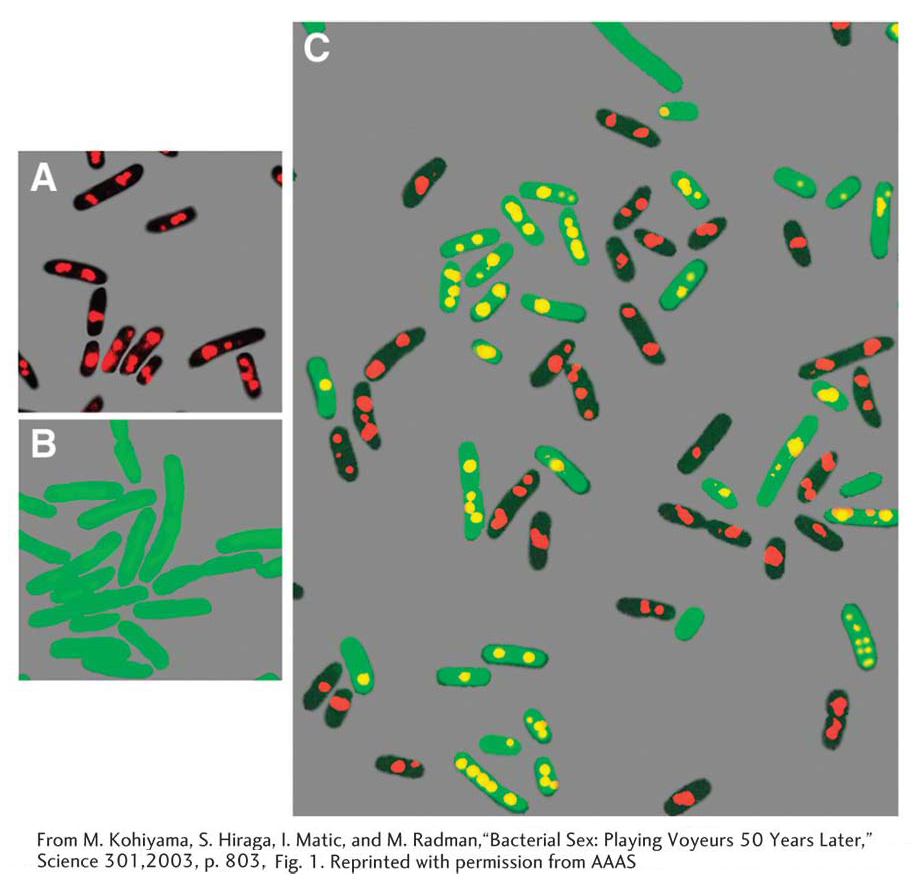
The photographs show a visualization of single-stranded DNA transfer in conjugating E. coli cells, with the use of special fluorescent antibodies. Parental Hfr strains (A) are black with red DNA. The red is from the binding of an antibody to a protein normally attached to DNA. The recipient F cells (B) are green due to the presence of the gene for a jellyfish protein that fluoresces green, and, because they are mutant for a certain gene, their DNA protein does not bind to antibody. When Hfr donor single-stranded DNA enters the recipient, it promotes atypical binding of this protein, which fluoresces yellow in this background. Part C shows Hfr’s (unchanged) and exconjugants (cells that have undergone conjugation) with yellow transferred DNA. A few unmated F cells are visible.
[From M. Kohiyama, S. Hiraga, I. Matic, and M. Radman, “Bacterial Sex: Playing Voyeurs 50 Years Later,” Science 301, 2003, p. 803, Fig. 1. Reprinted with permission from AAAS.]