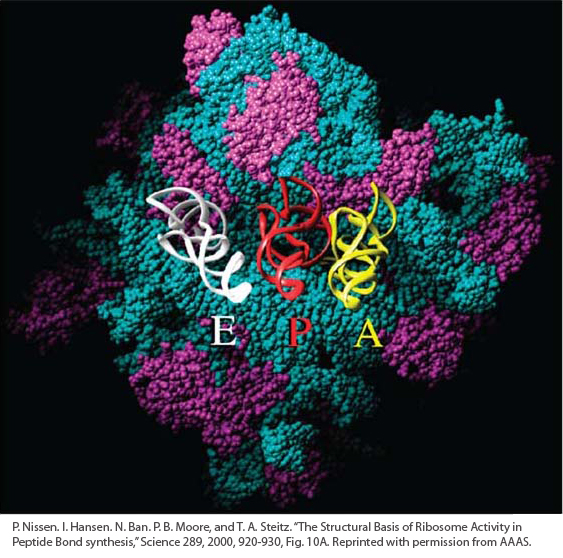
This image shows at atomic resolution a surface of the ribosome from the bacterium Haloarcula marismortui, deduced from X-ray crystallography. The part of the ribosome consisting of RNA is shown in blue; that consisting of protein is shown in purple. The white, red, and yellow structures in the center are tRNAs at the E, P, and A binding sites, their acceptor stems disappearing into a cleft in the ribosome.
[P. Nissen, J. Hansen, N. Ban, P. B. Moore, and T. A. Steitz, “The Structural Basis of Ribosome Activity in Peptide Bond Synthesis,” Science 289, 2000, 920-930, Fig. 10A. Reprinted with permission from AAAS.]