SUMMARY
In somatic cell division, the genome is transmitted by mitosis, a nuclear division. In this process, each chromosome replicates into a pair of chromatids and the chromatids are pulled apart to produce two identical daughter cells. (Mitosis can take place in diploid or haploid cells.) At meiosis, which takes place in the sexual cycle in meiocytes, each homolog replicates to form a dyad of chromatids; then, the dyads pair to form a tetrad, which segregates at each of the two cell divisions. The result is four haploid cells, or gametes. Meiosis can take place only in a diploid cell; hence, haploid organisms unite to form a diploid meiocyte.
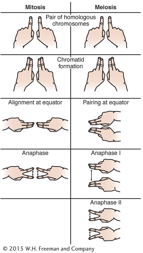
An easy way to remember the main events of meiosis, by using your fingers to represent chromosomes, is shown in Figure 2-33.
Genetic dissection of a biological property begins with a collection of mutants. Each mutant has to be tested to see if it is inherited as a single- A/A :
A/A :  A/a :
a/a is produced in the F2. (At the phenotypic level, this ratio is
A/a :
a/a is produced in the F2. (At the phenotypic level, this ratio is  A/– :
a/a.) The three single-
A and
a. The cellular basis of the equal segregation of alleles is the segregation of homologous chromosomes at meiosis. Haploid fungi can be used to show equal segregation at the level of a single meiosis (a 1:1 ratio in an ascus).
A/– :
a/a.) The three single-
A and
a. The cellular basis of the equal segregation of alleles is the segregation of homologous chromosomes at meiosis. Haploid fungi can be used to show equal segregation at the level of a single meiosis (a 1:1 ratio in an ascus).
71
The molecular basis for chromatid production in meiosis is DNA replication. Segregation at meiosis can be observed directly at the molecular (DNA) level. The molecular force of segregation is the depolymerization and subsequent shortening of microtubules that are attached to the centromeres. Recessive mutations are generally in genes that are haplosufficient, whereas dominant mutations are often due to gene haploinsufficiency.
In many organisms, sex is determined chromosomally, and, typically, XX is female and XY is male. Genes on the X chromosome (X-
Mendelian single-