9.3 tRNA: The Adapter

Once the genetic code was deciphered, scientists began to wonder how the sequence of amino acids of a protein was determined by the triplet codons of the mRNA. An early model, quickly dismissed as naive and unlikely, proposed that the mRNA codons could fold up and form 20 distinct cavities that directly bind specific amino acids in the correct order. Instead, in 1958, Crick recognized the following:

It is therefore a natural hypothesis that the amino acid is carried to the template by an adapter molecule, and that the adapter is the part which actually fits on to the RNA. In its simplest form [this hypothesis] would require twenty adapters, one for each amino acid.1

330

He speculated that the adapter “might contain nucleotides. This would enable them to join on the RNA template by the same ‘pairing’ of bases as is found in DNA.” Furthermore, “a separate enzyme would be required to join each adapter to its own amino acid.”

We now know that Crick’s “adapter hypothesis” is largely correct. Amino acids are in fact attached to an adapter (recall that adapters constitute a special class of stable RNAs called transfer RNAs). Each amino acid becomes attached to a specific tRNA, which then brings that amino acid to the ribosome, the molecular complex that will attach the amino acid to a growing polypeptide.

Codon translation by tRNA

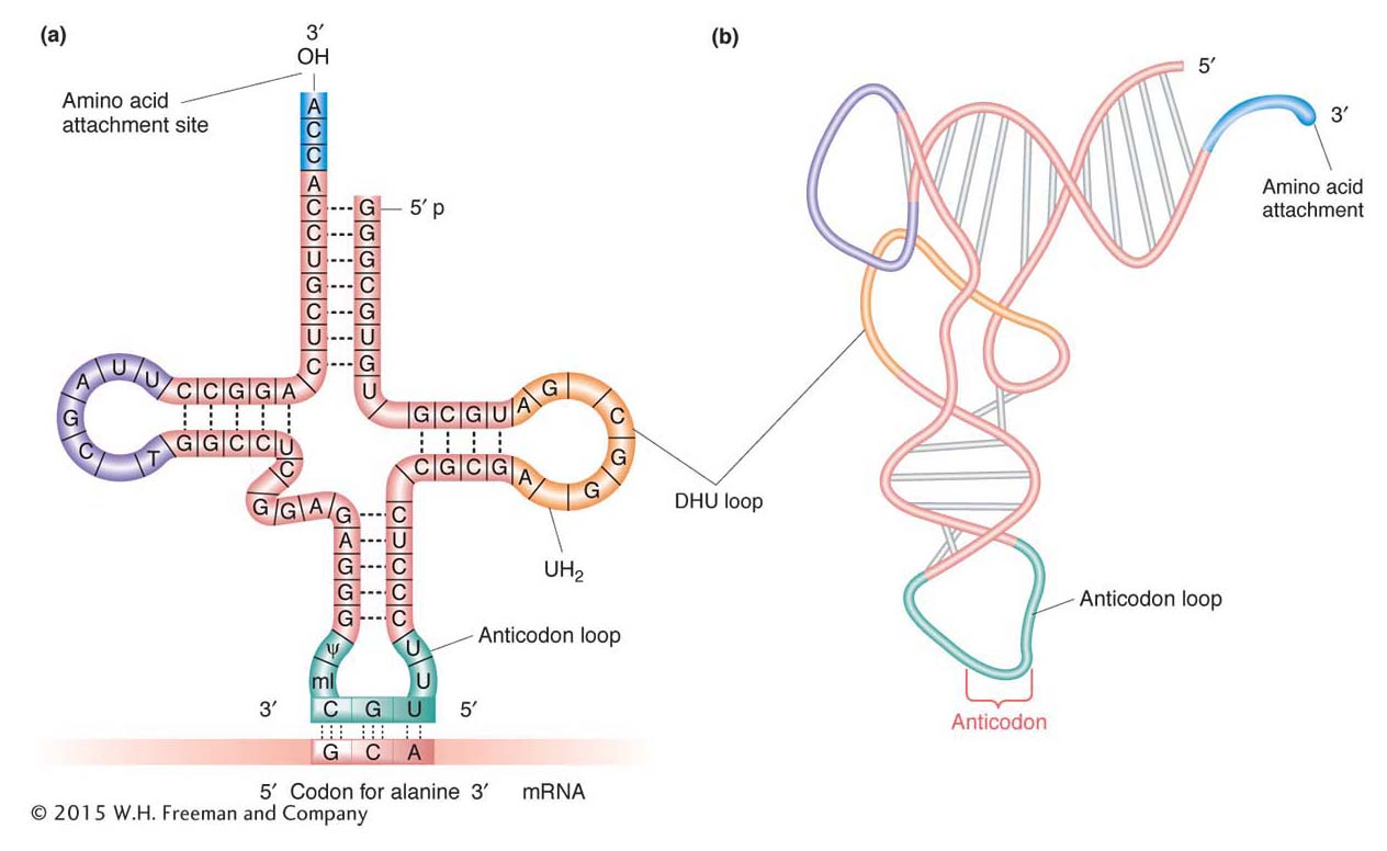
The structure of tRNA holds the secret of the specificity between an mRNA codon and the amino acid that it designates. The single-stranded tRNA molecule has a cloverleaf shape consisting of four double-helical stems and three single-stranded loops (Figure 9-6a). The middle loop of each tRNA is called the anticodon loop because it carries a nucleotide triplet called an anticodon. This sequence is complementary to the codon for the amino acid carried by the tRNA. The anticodon in tRNA and the codon in the mRNA bind by specific RNA-to-RNA base pairing. (Again, we see the principle of nucleic acid complementarity at work, this time in the binding of two different RNAs.) Because codons in mRNA are read in the 5′ × 3′ direction, anticodons are oriented and written in the 3′ × 5′ direction, as Figure 9-6a shows.

Figure 9-6: The structure of transfer RNA
Figure 9-6: (a) The structure of yeast alanine tRNA, showing the anticodon of the tRNA binding to its complementary codon in mRNA. (b) Diagram of the actual three-dimensional structure of yeast phenylalanine tRNA.
Figure 9-7: An aminoacyl-tMA synthetase attaches an amino acid to its tRNA
Figure 9-7: Each aminoacyl-tRNA synthetase has binding pockets for a specific amino acid and its cognate tRNA. By this means, an amino acid is covalently attached to the tRNA with the corresponding anticodon.

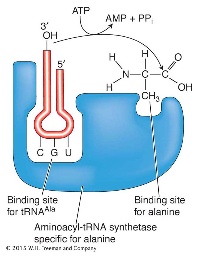
Amino acids are attached to tRNAs by enzymes called aminoacyl-tRNA synthetases. There are 20 of these enzymes in the cell, one for each of the 20 amino acids. Each amino acid has a specific synthetase that links it only to those tRNAs that recognize the codons for that particular amino acid. To catalyze this reaction, synthetases have two binding sites, one for the amino acid and the other for its cognate tRNA (Figure 9-7). An amino acid is attached at the free 3′ end of its tRNA, the amino acid alanine in the case shown in Figures 9-6a and 9-7. The tRNA with an attached amino acid is said to be charged.

331

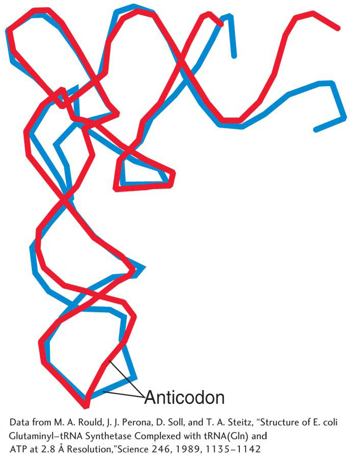
A tRNA normally exists as an L-shaped folded cloverleaf, as shown in Figure 9-6b, rather than the “flattened” cloverleaf shown in Figure 9-6a. The three-dimensional structure of tRNA was determined with the use of X-ray crystallography. In the years since this technique was used to deduce the double-helical structure of DNA, it has been refined so that it can now be used to determine the structure of very complex macromolecules such as the ribosome. Although tRNAs differ in their primary nucleotide sequence, all tRNAs fold into virtually the same L-shaped conformation except for differences in the anticodon loop and aminoacyl end. This similarity of structure can be easily seen in Figure 9-8, which shows two different tRNAs superimposed. Conservation of structure tells us that shape is important for tRNA function.

Figure 9-8: Two superimposed tRNAs
Figure 9-8: When folded into their correct three-dimensional structures, the yeast tRNA for glutamine (blue) almost completely overlaps the yeast tRNA for phenylalanine (red) except for the anticodon loop and aminoacyl end.
[Data from M. A. Rould, J. J. Perona, D. Soll, and T. A. Steitz, “Structure of E. coli Glutaminyl-tRNA Synthetase Complexed with tRNA(Gln) and ATP at 2.8 A Resolution,” Science 246, 1989, 1135-1142.]

What would happen if the wrong amino acid were covalently attached to a tRNA? A convincing experiment answered this question. The experiment used cysteinyl-tRNA (tRNACys), the tRNA specific for cysteine. This tRNA was “charged” with cysteine, meaning that cysteine was attached to the tRNA. The charged tRNA was treated with nickel hydride, which converted the cysteine (while still bound to tRNACys) into another amino acid, alanine, without affecting the tRNA:

Protein synthesized with this hybrid species had alanine wherever we would expect cysteine. The experiment demonstrated that the amino acids are “illiterate”; they are inserted at the proper position because the tRNA “adapters” recognize the mRNA codons and insert their attached amino acids appropriately. Thus, the attachment of the correct amino acid to its cognate tRNA is a critical step in ensuring that a protein is synthesized correctly. If the wrong amino acid is attached, there is no way to prevent it from being incorporated into a growing protein chain.

Degeneracy revisited

As can be seen in Figure 9-5, the number of codons for a single amino acid varies, ranging from one codon (UGG for tryptophan) to as many as six (UCC, UCU, UCA, UCG, AGC, or AGU for serine). Why the genetic code contains this variation is not exactly clear, but two facts account for it:

  1. Most amino acids can be brought to the ribosome by several alternative tRNA types. Each type has a different anticodon that base-
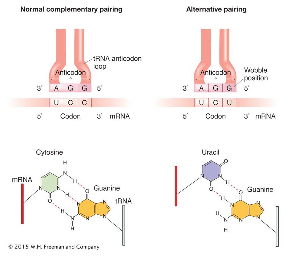
pairs with a different codon in the mRNA.

  2. Certain charged tRNA species can bring their specific amino acids to any one of several codons. These tRNAs recognize and bind to several alternative codons, not just the one with a complementary sequence, through a loose kind of base pairing at the 3′ end of the codon and the 5′ end of the anticodon. This loose pairing is called wobble.

Wobble is a situation in which the third nucleotide of an anticodon (at the 5 ′ end) can form either of two alignments (Figure 9-9). This third nucleotide can form hydrogen bonds either with its normal complementary nucleotide in the third position of the codon or with a different nucleotide in that position.

Figure 9-9: Wobble allowsto recognize two codons
Figure 9-9: In the third site (5′ end) of the anticodon, G can take either of two wobble positions, thus being able to pair with either U or C. This ability means that a single tRNA species carrying an amino acid (in this case, serine) can recognize two codons—UCU and UCC—in the mRNA.

“Wobble rules” dictate which nucleotides can and cannot form hydrogen bonds with alternative nucleotides through wobble (Table 9-1). In Table 9-1, the letter I stands for inosine, one of the rare bases found in tRNA, often in the anticodon.

5′ end of anticodon

3′ end of codon

G

C or U

C

G only

A

U only

U

A or G

I

U, C, or A

Table 9-1: Codon-Anticodon Pairings Allowed by the Wobble Rules

332

KEY CONCEPT

The genetic code is said to be degenerate because, in many cases, more than one codon is assigned to a single amino acid; in addition, several codons can pair with more than one anticodon (wobble).