Chapter Introduction
Large-
617
Large-
Chromosomal
Changes
CHAPTER
17
LEARNING OUTCOMES
After completing this chapter, you will be able to
Distinguish between the major types of chromosomal mutations at the cytological level.
Draw meiotic pairing configurations for all the major chromosomal mutations.
Predict progeny ratios of specific autopolyploids heterozygous for one or more genes.*
Design crosses to synthesize an allotetraploid.
Predict the outcome of first and second division meiotic nondisjunction.*
Identify an aneuploid using genetic criteria.*
Predict ratios in progeny of specific aneuploids.*
Distinguish between the main human aneuploid types.
In progeny analysis, diagnose the presence of one of the major types of chromosome rearrangements (translocations, inversions, deletions, duplications).*
In a cross involving a known specific rearrangement, predict the inheritance of genes linked and unlinked to the rearrangement.*
Predict patterns of expression of genes potentially affected by position-
effect variegation.
*In the case of chromosomal mutations, progeny analysis involves analyzing patterns of one or more of the following: sterility, lethality, and phenotypic proportions of genes heterozygous in the crosses.
OUTLINE
17.1 Changes in chromosome number
17.2 Changes in chromosome structure
17.3 Overall incidence of human chromosome mutations
618
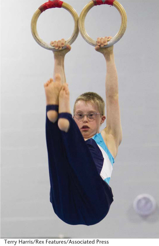
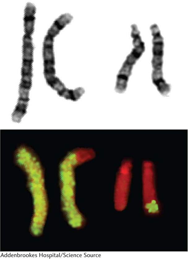
A young couple is planning to have children. The husband knows that his grandmother had a child with Down syndrome by a second marriage. Down syndrome is a set of physical and mental disorders caused by the presence of an extra chromosome 21 (Figure 17-1). No records of the birth, which occurred early in the twentieth century, are available, but the couple knows of no other cases of Down syndrome in their families.
The couple has heard that Down syndrome results from a rare chance mistake in egg production and therefore decide that they stand only a low chance of having such a child. They decide to have children. Their first child is unaffected, but the next conception aborts spontaneously (a miscarriage), and their second child is born with Down syndrome. Was their having a Down syndrome child a coincidence, or did a connection between the genetic makeup of the child’s father and that of his grandmother lead to their both having Down syndrome children? Was the spontaneous abortion significant? What tests might be necessary to investigate this situation? The analysis of such questions is the topic of this chapter.
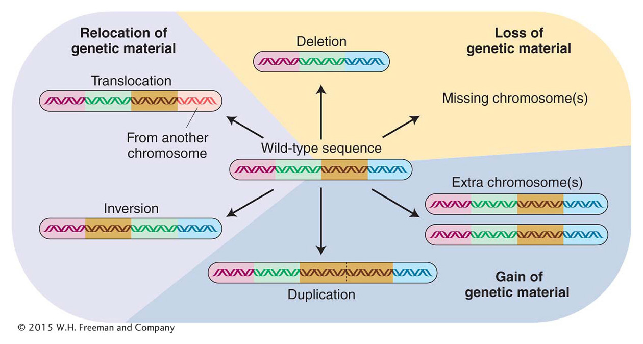
We have seen throughout the book that gene mutations are an important source of change in the genomic sequence. However, the genome can also be remodeled on a larger scale by alterations to chromosome structure or by changes in the number of copies of chromosomes in a cell. These large-
Chromosome mutations are important from several biological perspectives. First, they can be sources of insight into how genes act in concert on a genomic scale. Second, they reveal several important features of meiosis and chromosome architecture. Third, they constitute useful tools for experimental genomic manipulation. Fourth, they are sources of insight into evolutionary processes. Fifth, chromosomal mutations are regularly found in humans, and some of these mutations cause genetic disease.
Many chromosome mutations cause abnormalities in cell and organismal function. Most of these abnormalities stem from changes in gene number or gene position. In some cases, a chromosome mutation results from chromosome breakage. If the break occurs within a gene, the result is functional disruption of that gene.
For our purposes, we will divide chromosome mutations into two groups: changes in chromosome number and changes in chromosome structure. These two groups represent two fundamentally different kinds of events. Changes in chromosome number are not associated with structural alterations of any of the DNA molecules of the cell. Rather, it is the number of these DNA molecules that is changed, and this change in number is the basis of their genetic effects. Changes in chromosome structure, on the other hand, result in novel sequence arrangements within one or more DNA double helices. These two types of chromosome mutations are illustrated in Figure 17-2, which is a summary of the topics of this chapter. We begin by exploring the nature and consequences of changes in chromosome number.