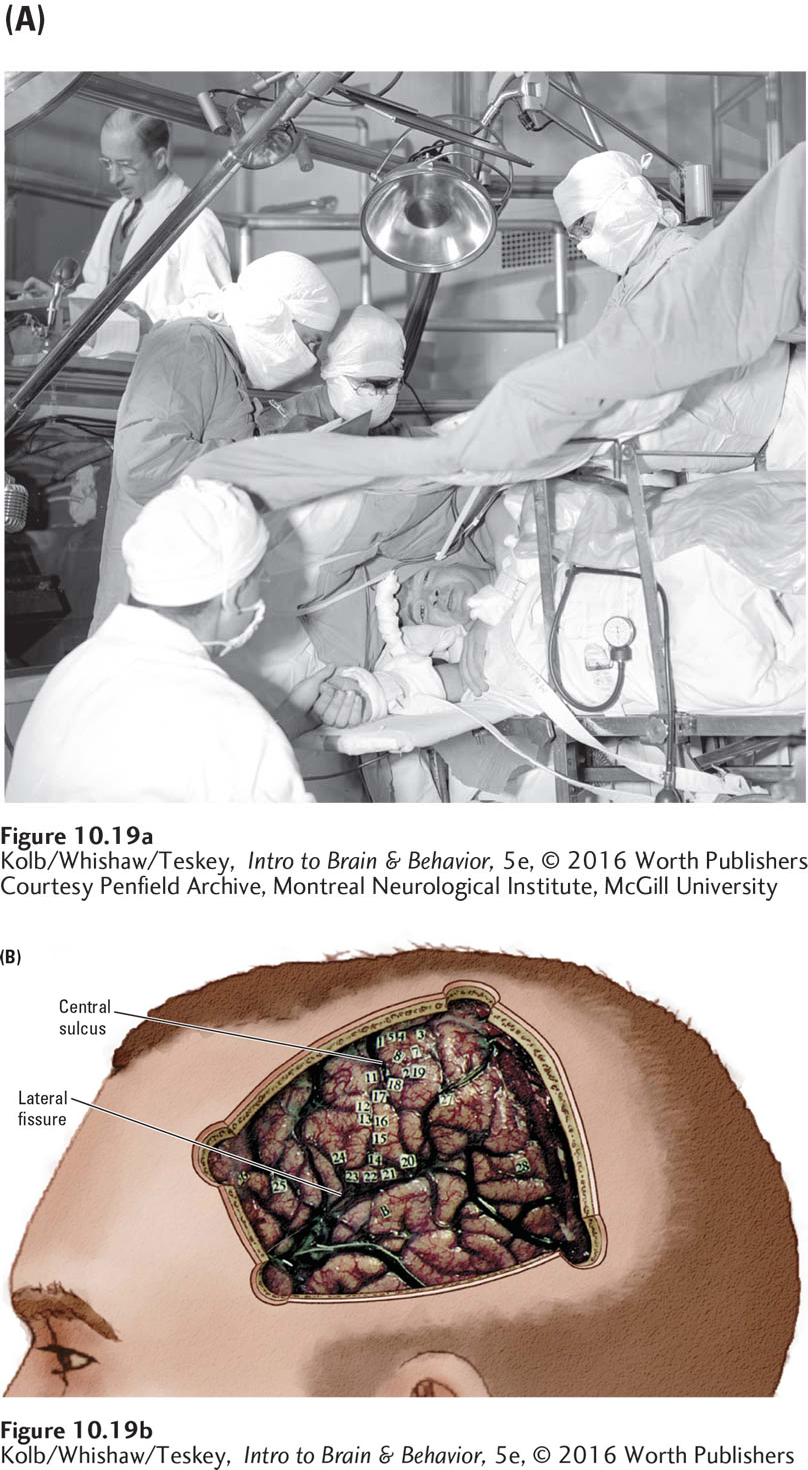
image
FIGURE 10-19 Mapping Cortical Functions (A) Neurosurgery for eligible epilepsy patients who failed to respond to antiseizure medications. The patient is fully conscious, lying on his right side, and kept comfortable with local anesthesia. Wilder Penfield stimulates discrete cortical areas in the patient’s exposed left hemisphere. In the background, a neurologist monitors an EEG recorded from each stimulated area to help identify the epileptogenic focus. The anesthetist (seated) observes the patient’s responses to the cortical stimulation. (B) A drawing overlies a photograph of the patient’s exposed brain. The numbered tickets identify points Penfield stimulated to map the cortex in this patient’s brain. At points 26, 27, and 28, a stimulating electrode disrupted speech. Point 26 presumably is in Broca’s area, 27 is the motor cortex facial control area, and 28 is in Wernicke’s area.
Courtesy Penfield Archive, Montreal Neurological Institute, McGill University