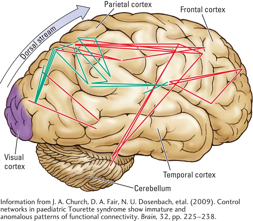
In fMRI analyses of young adults with Tourette syndrome, brain areas that show enhanced (green) or decreased (red) connectivity suggest abnormalities in dorsal stream structures linking the parietal cortex to the frontal cortex.
Information from J. A. Church, D. A. Fair, N. U. Dosenbach, et al. (2009). Control networks in paediatric Tourette syndrome show immature and anomalous patterns of functional connectivity. Brain, 32, pp. 225–