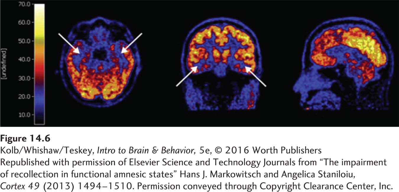
image
FIGURE 14-6 Lost Episodes Left to right: Horizontal, frontal, and sagittal sections imaged in a patient with impaired autobiographical memory. White arrows point to areas of reduced glucose metabolism in frontal and temporal regions as she attempts to retrieve remote personal memories. Her MRI scan was normal.
Republished with permission of Elsevier Science and Technology Journals from “The impairment of recollection in functional amnesic states” Hans J. Markowitsch and Angelica Staniloiu, Cortex 49 (2013) 1494–1510. Permission conveyed through Copyright Clearance Center, Inc.