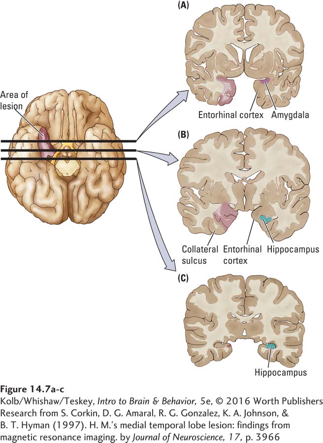
image
FIGURE 14-7 Extent of H. M.’s Surgery H. M.’s right-hemisphere lesion is highlighted in the brain viewed ventrally. The lesion runs along the wall of the medial temporal lobe. The left side of the brain has been left intact to show the relative location of the medial temporal structures. Parts A, B, and C, based on MRI scans, depict a series of coronal sections of H. M.’s brain.
Research from S. Corkin, D. G. Amaral, R. G. Gonzalez , K. A. Johnson, & B. T. Hyman (1997). H. M.’s medial temporal lobe lesion: findings from magnetic resonance imaging. by Journal of Neuroscience, 17, p. 3966.