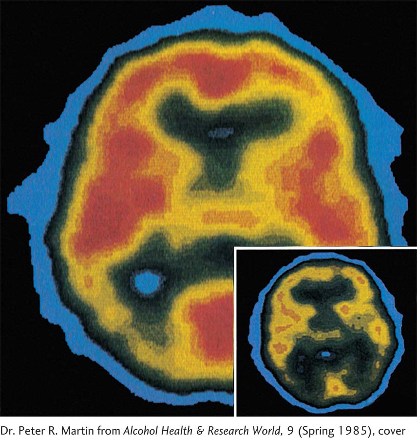
PET scans from a healthy patient (larger image) and a Korsakoff patient (inset) reveal reduced activity in the frontal lobes of the diseased brain. (The frontal lobes are at the bottom center of each scan.) Red and yellow represent areas of high metabolic activity; activity is lower in the darker areas.
Dr. Peter R. Martin from Alcohol Health & Research World, 9 (Spring 1985), cover