image
FIGURE 15-9 Parcellation of Cerebral Cortical Networks An estimate of 17 cortical networks based on fcMRI data from 1000 participants. Each color represents a network. Some, such as the blue auditory areas in temporal lobe, are localized; others are widely distributed, such as the yellow regions, which reveal prefrontal–posterior parietal connectivity.
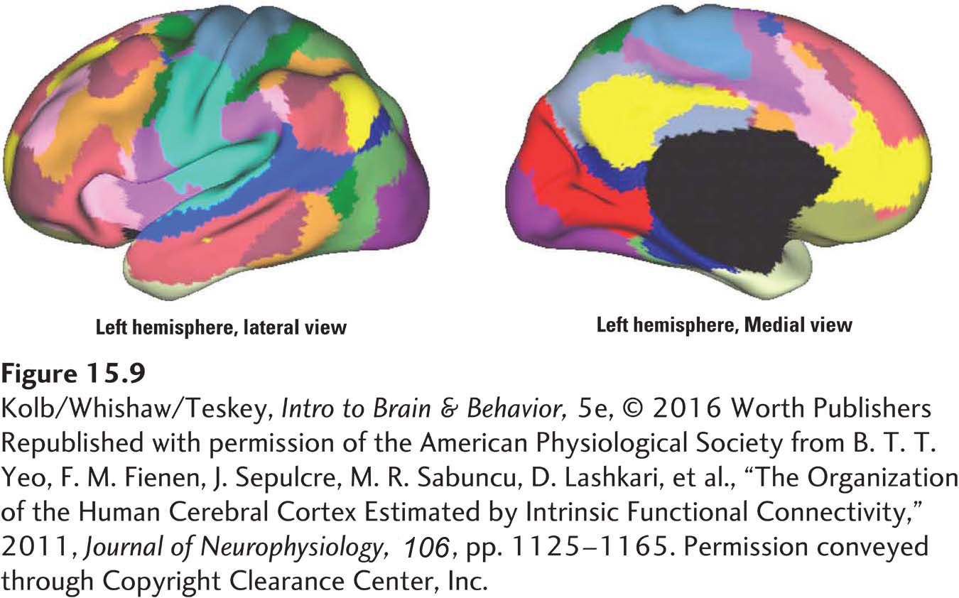
Republished with permission of the American Physiological Society from B. T. T. Yeo, F. M. Fienen, J. Sepulcre, M. R. Sabuncu, D. Lashkari, et al., “The Organization of the Human Cerebral Cortex Estimated by Intrinsic Functional Connectivity,” 2011, Journal of Neurophysiology, 106, pp. 1125–1165. Permission conveyed through Copyright Clearance Center, Inc.