image
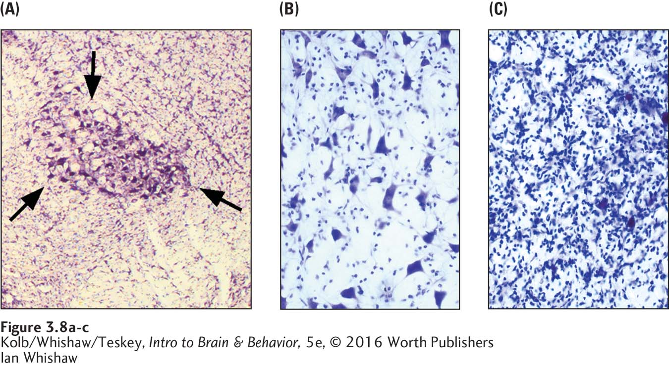
FIGURE 3-8 Detecting Brain Damage (A) Arrows indicate the red nucleus in a rat brain. (B) Close-up of cresyl violet–stained neurons, the large dark bodies, in the healthy red nucleus. (C) After exposure to a neurotoxin, only microglia, the small dark objects in the micrograph, survive.
Ian Whishaw