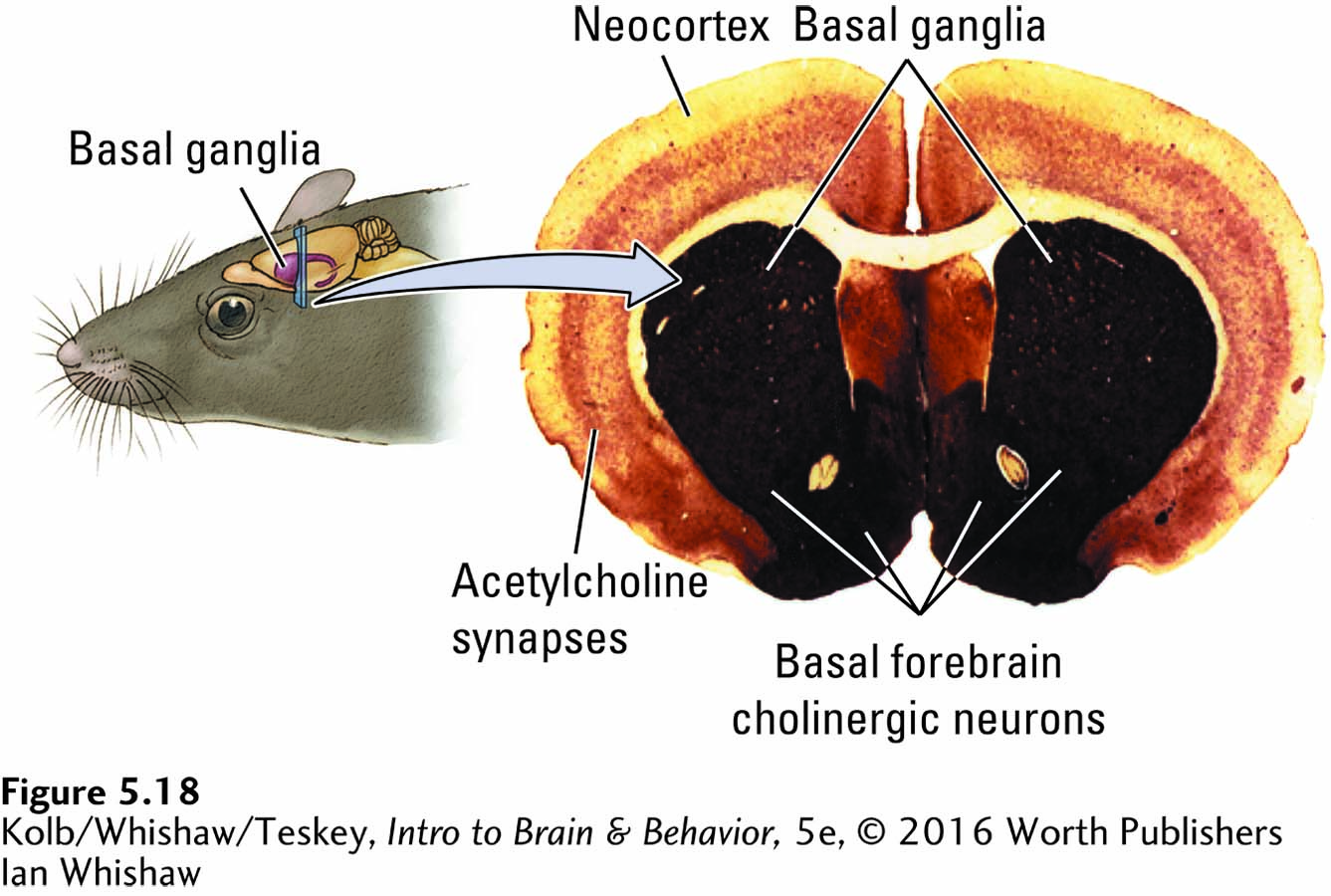
image
FIGURE 5-18 Cholinergic Activation Drawing at left shows the cortical location of the micrograph at right, stained to reveal AChE. Cholinergic neurons in the rat’s basal forebrain project to the neocortex, and the darkly stained bands in the cortex show areas rich in cholinergic synapses. The darker central parts of the section, also rich in cholinergic neurons, are the basal ganglia.
Ian Whishaw