image
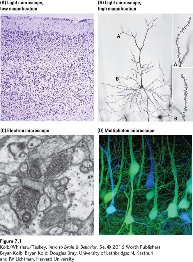
FIGURE 7-1 Staining Cerebral Neurons Viewed through a light microscope, (A) Nissl-stained section of parietal cortex shows all cell bodies but no cell processes (axons and dendrites). (B) At higher magnification, an individual Golgi-stained pyramidal cell from the parietal cortex is visible. The cell body (dark triangular shape at center) and spiny dendrites (A and B) are visible in detail at right. (C) The view through an electron microscope shows neuronal synapses in detail. (D) Multiple images from a multiphoton microscope, merged to generate a three-dimensional image of living tissue.
Bryan Kolb
Bryan Kolb
Douglas Bray, University of Lethbridge
N. Kasthuri and JW Lichtman, Harvard University