image
FIGURE 7-13 CT Scan and Brain Reconstruction (A) Dorsal view of a horizontal CT scan of a subject with Broca’s aphasia. The dark region at the left anterior is the area of the lesion. (B) A schematic representation of the horizontal section, with the area of the lesion shown in blue. (C) A reconstruction of the brain, showing a lateral view of the left hemisphere with the lesion shown in blue.
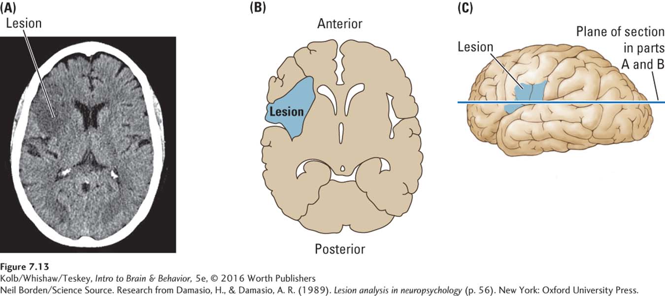
Research from Damasio, H., & Damasio, A. R. (1989). Lesion analysis in neuropsychology (p. 56). New York: Oxford University Press.
Neil Borden/Science Source