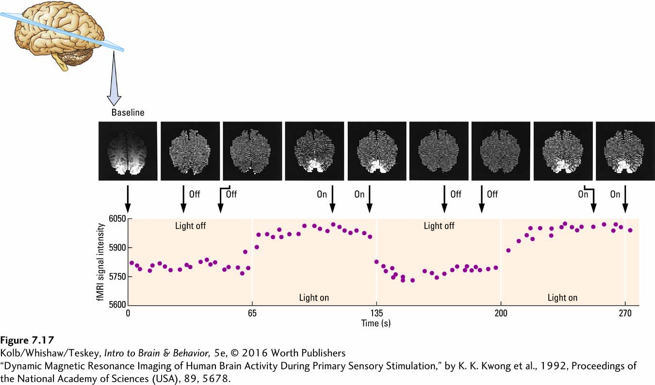
image
FIGURE 7-17 Imaging Changes in Brain Activity Functional MRI sequence of a horizontal section at mid-occipital lobe (bottom of each image) in a normal human brain during visual stimulation. A baseline acquired in darkness (far left) was subtracted from the subsequent images. The participant wore tightly fitting goggles containing light-emitting diodes that were turned on and off as a rapid sequence of scans was obtained over 270 seconds. Note the prominent activity in the visual cortex when the light is on and the rapid cessation of activity when the light is off, all measured in the graph of signal intensity below the images.
“Dynamic Magnetic Resonance Imaging of Human Brain Activity During Primary Sensory Stimulation,” by K. K. Kwong et al., 1992, Proceedings of the National Academy of Sciences (USA), 89, 5678.