image
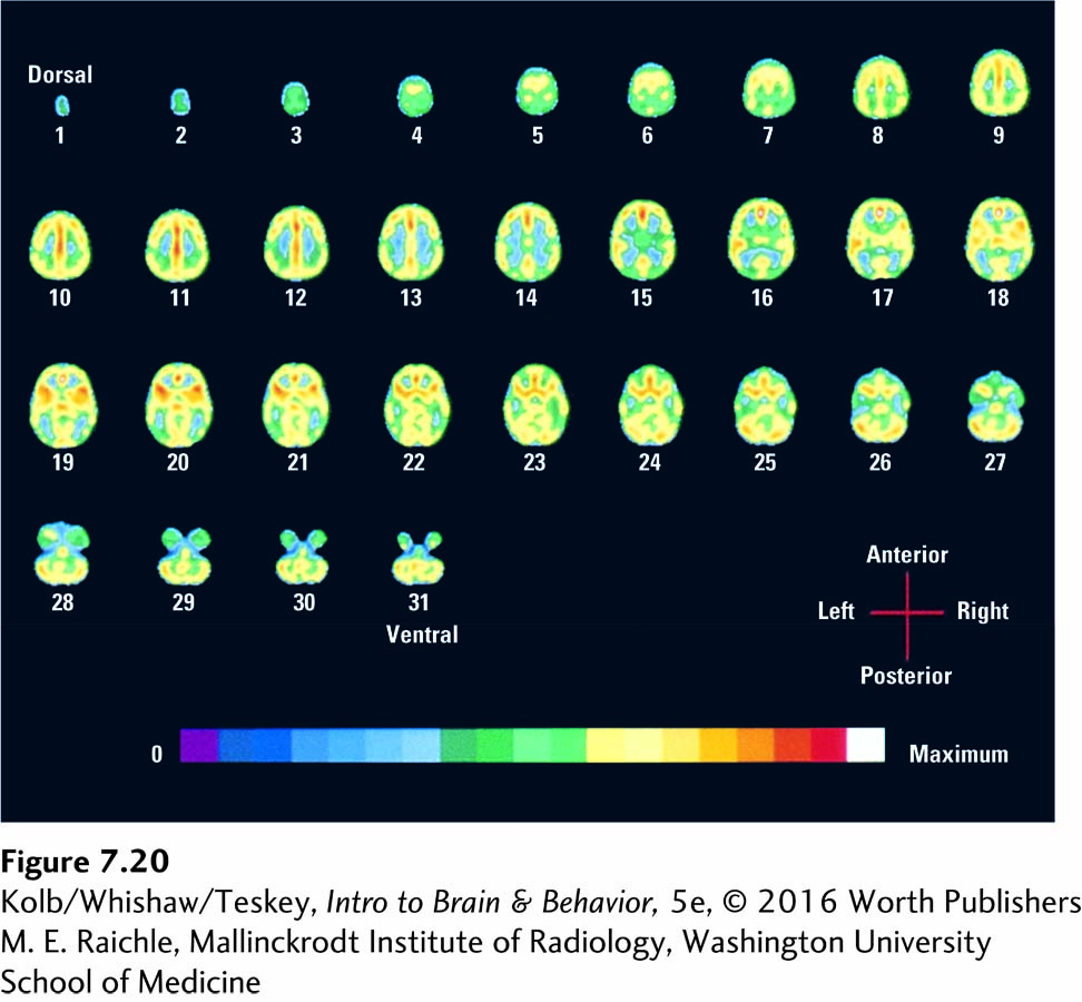
FIGURE 7-20 Resting State PET images of blood flow obtained while a single subject rested quietly with eyes closed. Each scan represents a horizontal brain section, from the dorsal surface (1) to the ventral surface (31). Development of rs-fMRI suggests that resting-state PET analysis may emerge.
M. E. Raichle, Mallinckrodt Institute of Radiology, Washington University School of Medicine