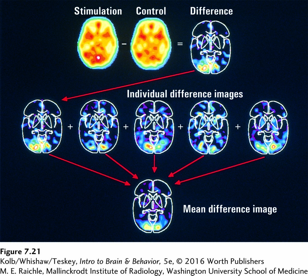
image
FIGURE 7-21 The Procedure of Subtraction In the upper row of scans, the control condition, resting while looking at a static fixation point (control), is subtracted from the experimental condition, looking at a flickering checkerboard (stimulation). The subtraction produces a different scan for each of five experimental subjects, shown in the middle row, but all show increased blood flow in the occipital region. The difference scans are averaged to produce the representative image at the bottom.
M. E. Raichle, Mallinckrodt Institute of Radiology, Washington University School of Medicine