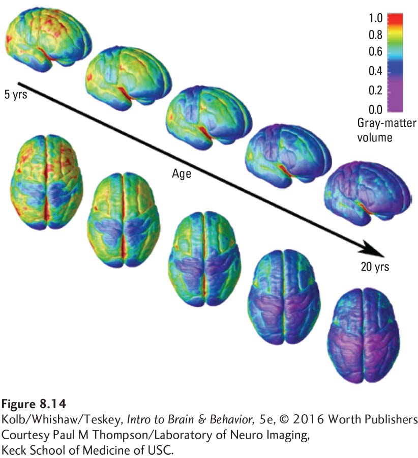
image
FIGURE 8-14 Progressive Changes in Cortical Thickness MRI scans track the maturation of gray matter in typical development, revealing the length and pattern of maturation from the back of the cortex to the front. Cortical thinning and increased surface area progress together.
Courtesy Paul M Thompson/Laboratory of Neuro Imaging, Keck School of Medicine of USC.