15-3 Expanding Frontiers of Cognitive Neuroscience
Sophisticated noninvasive stimulation and recording techniques for measuring the brain’s electrical activity and noninvasive brain imaging methods led to a major shift in studying brain and behavior: cognitive neuroscience, the field that studies the neural bases of cognition. Cognitive neuroscience focuses on high-
CLINICAL FOCUS 15-4
Neuropsychological Assessment
In this high-
To illustrate the nature and power of neuropsychological assessment, we compare three patients’ test performance on an array of tests selected from among those used in a complete neuropsychological assessment. The five tests presented here measure verbal and visual memory, verbal fluency, abstract reasoning, and reading. Performance was compared with that of a healthy control participant.
In the delayed memory tests—
Half an hour later, without warning, they were asked to perform the tasks again. Their performance on these tests yielded the delayed verbal and visual memory scores listed in the table.
| Subjects’ Scores | ||||
|---|---|---|---|---|
| Test | Control | J. N. | E. B. | J. W. |
| Delayed verbal memory | 17 | 9* | 16 | 16 |
| Delayed visual memory | 12 | 14 | 8* | 12 |
| Verbal fluency | 62 | 62 | 66 | 35* |
| Card- |
9 | 10 | 12 | 56* |
| Reading | 15 | 21 | 22 | 17 |
|
*Atypically poor score. |
||||
In the delayed verbal fluency test, patients and control had 5 minutes to write down as many words as they could think of that start with the letter s, excluding numbers and people’s names. Then came the Wisconsin Card Sorting Test, which assesses abstract reasoning (see Figure 15-8). Finally, all were given a reading test.
The first patient, J. N., was a 28-
When we saw J. N. a year after surgery that successfully removed the tumor, he had returned to his job as a personnel manager. His intelligence was still superior, but as the score summary shows, he was impaired on the delayed verbal memory test, recalling only about 50 percent as much as the control and other subjects.
The second patient, E. B., was a college senior majoring in psychology. An aneurysm in her right temporal lobe had burst due to a bulge in the artery. The anterior part of that lobe had been removed. E. B. was of above-
The third patient, also of above-
We saw J. W. 10 years after his surgery. He was still on the police force but working a desk job. His verbal fluency was markedly reduced, as was his ability to solve the card-
Two principles emerge from the results of these three neuropsychological assessments:
Brain functions are localized. Damage to different brain regions produces different symptoms.
Brain organization is asymmetrical. Left-
hemisphere damage preferentially affects verbal functions; right- hemisphere damage preferentially affects nonverbal functions.
534
Sophisticated imaging techniques are helping cognitive neuroscientists map the human brain. Its methods assist social psychologists in discovering how the brain mediates social interactions and economists in discovering how the brain makes decisions.
Mapping the Brain
Among the great scientific challenges of the twenty-
For more on the HCP, including connectome images, go to www.humanconnectomeproject.org. National Geographic published an accessible article in February 2014.
The HCP’s goal is to provide a reference atlas for those seeking to understand human brain function and dysfunction. It has attracted considerable public interest, and an explosion of studies outside the HCP also seeks to map the human brain. For example, in a review, Roser Sala-
Using fMRI in Brain Mapping
The fcMRI technique uses resting-
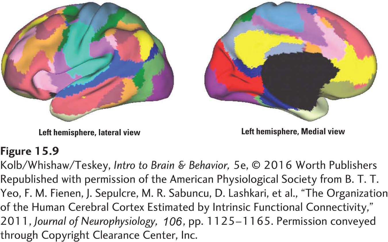
The cerebral cortex is made up of primary sensory and motor networks as well as the multiple, large-
In contrast, the association networks include areas distributed throughout the prefrontal, parietal, anterior temporal, and midline regions. In Figure 15-9, the distributed yellow regions show prefrontal–
Unlike DTI, fcMRI does not measure static anatomical connectivity but rather uses temporal (time-
535
Tractography Using Diffusion Tensor Imaging
DTI studies provide results, often called tractography, that complement the networks mapped by fcMRI. Tractography measures actual neuroanatomical pathways that can be related to specific traits. Traditional postmortem tract tracing was performed on single brains. Today, tractography can be performed quickly on many living brains, and measurements can be made simultaneously in the entire brain. This advance allows researchers to correlate specific behavioral traits with specific patterns of connectivity.
Psyche Loui and her colleagues (2011) were interested in the neural basis of perfect (or absolute) pitch, the ability not only to discriminate among musical notes but also to name any note heard. Perfect pitch is rare among humans but shared by people with remarkable musical talents. Think Mozart. Development of perfect pitch is sensitive to early experiences, including musical training and exposure to tonal languages such as Japanese.
Loui’s team studied musicians, with and without absolute pitch, who were matched in gender, age, handedness, ethnicity, IQ score, and years of musical training. Participants were given a test of pitch-
The investigators hypothesized that absolute pitch could be related to increased connectivity in brain regions that process sounds. They used DTI to reconstruct white-
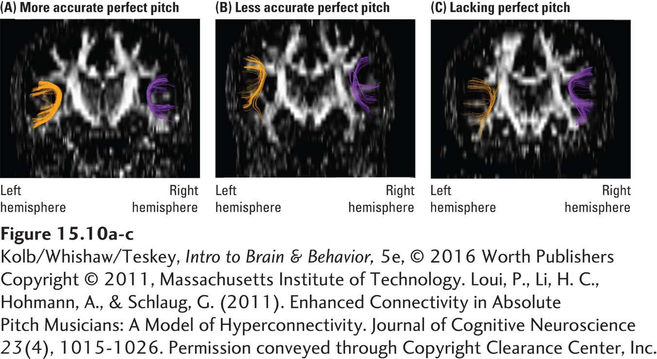
Although enhanced connectivity appears in both hemispheres of the musicians, when the investigators correlated performance on a test of absolute pitch with tract volume, only left-
It is tempting to speculate that other exceptional talents, such as creativity, might be related to hyperconnectivity in cerebral regions. Conversely, we can speculate that reduced structural and functional connectivity is related to cognitive impairments after acquired brain injuries and/or neurodevelopmental and psychiatric disorders.
Cognition and the Cerebellum
Research Focus 2-1 describes the effects of cerebellar agenesis.
So far we have emphasized the role of the neocortex in cognitive functions, in part because of its marked expansion in size and neuron numbers in primate brains. Yet the human cerebellum accounts for 80 percent of the brain’s neurons. The cerebellum has long been known to play a central role in motor control and motor learning, but the concurrent evolution of neocortex, cerebellum, and cognitive complexity in primates suggests that the cerebellum may play a larger role in cognitive processes than investigators have appreciated (e.g., Barton, 2012).
536
The extensive neocortex–
Social Neuroscience
By combining cognitive neuroscience tools, especially functional neuroimaging, with abstract constructs from social psychology, social neuroscience seeks to understand how the brain mediates social interactions. Matthew Lieberman (2007) identified broad themes that attempt to encompass all cognitive processes involved in understanding and interacting with others and in understanding ourselves.
Understanding Others
Animals’ mind and experiences are not open to direct inspection. We infer animals’ mind in part by observing their behaviors and peoples’ mind by listening to their words. In doing so, we may develop a theory of mind, the attribution of mental states to others. Theory of mind includes an understanding that others may have feelings and beliefs different from our own. This broader understanding has led some investigators to conclude that theory of mind may be uniquely human. But many researchers who study apes strongly believe that other apes, too, possess a theory of mind.
Many fMRI studies over the past decade suggest that the brain region believed most closely associated with theory of mind is the dorsolateral prefrontal cortex (see Figure 15-2). Human prefrontal regions are disproportionately large as corrected for brain size, but other apes also have large prefrontal regions. The anatomy supports the likelihood that they also possess a theory of mind.
The capacity to understand others can also be inferred from the presence of empathy. For example, when participants watch videos of others smelling disgusting odors, they report a feeling of disgust. Lieberman and his colleagues (Rameson et al., 2012) used fMRI to assess the neural correlates of empathy by asking participants to empathize with sad images. Empathy correlated with increased activity in the medial prefrontal region, suggesting that the area is critical for empathic experience.
Understanding Oneself
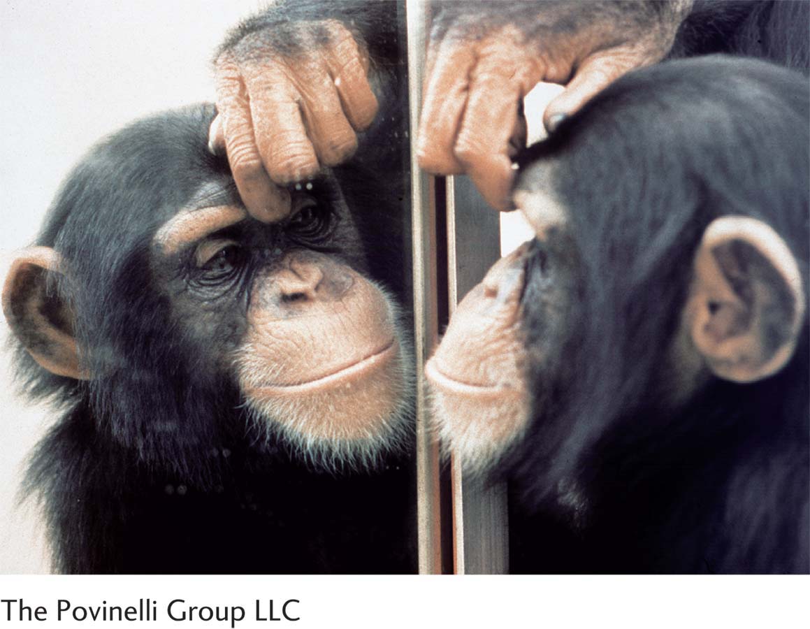
Not only are we humans aware of others’ intentions, we also have a sense of self. Humans and apes have the ability to recognize themselves in a mirror, an ability that human infants demonstrate by about 21 months of age. Studies using fMRI show that when we recognize our own face versus the face of familiar others, brain activity increases in the right lateral prefrontal cortex and in the lateral parietal cortex. The parietal cortex activation is thought to reflect the body’s recognition of what itself feels like.
537
But self-
Self-
Self-
Section 8-2 describes unique aspects of frontal lobe development that extend beyond childhood—
Children are often poor self-
Feeling and treating physical pain is a topic in Section 11-4. Focus 12-1 reports on emotional pain.
Not only can humans control their emotions; they also have expectations about how a stimulus might feel (e.g., an injection by syringe). Our expectations can alter the actual feeling of an event. It is common for people to say ouch when they do something like stub a toe, even if they actually feel no pain. Nobukatsu Sawamoto and colleagues (2000) found that when participants expect pain, activity increases in the anterior cingulate cortex (see Figure 15-2B), a region associated with pain perception, even if the stimulus turns out not to be painful.
Living in a Social World
We spend much of our waking time interacting with others socially. In a sense, our understanding of our self and our social interactions link together into a single mental action. One important aspect of this behavior includes forming attitudes and beliefs about ourselves and about others. When we express attitudes (including prejudices) toward ideas or human groups, brain imaging shows activation in prefrontal, anterior cingulate, and lateral parietal regions.
Samuel McLure and colleagues (2004) took advantage of the fact that many people have strong attitudes toward cola-
As a group, participants failed to discriminate the drinks accurately when they were presented in a blind taste test. Brain activity was also equivalent for each cola in the blind condition. However, when participants believed that they were drinking Coca-
Social Cognition and Brain Activity
Social cognitions running the gamut from understanding ourselves to understanding others clearly are associated with activation of specific brain regions, especially prefrontal regions. The obvious conclusion is that prefrontal activity produces our social cognitions, just as activity in visual regions produces our visual perceptions. But this conclusion has proved controversial. Ed Vul and colleagues (2009, 2012) go so far as to suggest that “correlations in social neuroscience are voodoo.” Their assertion has led to strong disputations (e.g., Lieberman et al., 2012). The arguments are complex, focus on the nature of the analysis of fMRI data, and will certainly continue. In our view, the debate does not impugn the general conclusion that brain states produce behavioral states.
Neuroeconomics
Leonard Mlodinow’s wonderful 2009 book, The Drunkard’s Walk: How Randomness Rules Our Lives, offers many everyday examples.
538
Historically, economics was a discipline based on the “rational actor,” the belief that people make rational decisions. In the real world, people often make decisions based on assumption or intuition, as is common in gambling. Why don’t people always make rational decisions?
The cerebral processes underlying human decision making are not easily inferred from behavioral studies. But investigators in the field of neuroeconomics, which combines ideas from economics, psychology, and neuroscience, attempt to explain those processes by studying patterns of brain activity as people make decisions in real time. The general assumption among neuroeconomists is that two neural decision pathways influence our choices. One is deliberate, slow, rule-
Figure 6-17 maps the dopaminergic pathways associated with reward.
If people must make quick decisions they believe will provide immediate gain, widespread activity appears in the dopaminergic reward system. This includes the ventromedial prefrontal cortex and ventral striatum (nucleus accumbens): the reflexive pathway. If slower, deliberative decisions are possible, activity is greater in the lateral prefrontal, medial temporal, and posterior parietal cortex, the areas that form the reflective pathway.
Neuroeconomists are looking to identify patterns of neural activity in everyday decision making, patterns that may help account for how people make decisions about their finances, social relations, and other personal choices. Although most neuroeconomic studies to date have used fMRI, in principle these studies could also use other noninvasive imaging technologies. Epigenetic factors probably contribute to developing the balance between the reflective and reflexive systems in individuals. Epigenetic studies therefore may help explain why many people make decisions that are not in their long-
Because of predictions that all mammals will have similar reflective and reflexive decision systems and because all animals make decisions, the neural bases of decision processes in nonhumans undoubtedly will receive more study in the future.
15-3 REVIEW
Expanding Frontiers of Cognitive Neuroscience
Before you continue, check your understanding.
Question 1
Noninvasive imaging techniques enable cognitive psychologists to investigate the neural bases of thought in the “normal” brain, leading to the field called ____________.
Question 2
Imaging methods such as DTI and fcMRI are allowing researchers to develop a ____________, a map of the complete structural and functional fiber pathway connections in the living human brain.
Question 3
The role of the ____________ in cognition, formerly unappreciated, is now attracting researchers’ attention.
Question 4
Social neuroscience is an interdisciplinary field that seeks to understand how the brain mediates ____________.
Question 5
Our attribution of mental states to others is known as ____________.
Question 6
Neuroeconomics seeks to understand the neural bases of ____________.
Question 7
List four general themes of social neuroscience research.
Answers appear in the Self Test section of the book.