2-1 Overview of Brain Function and Structure
35
The brain’s primary function is to produce behavior, or movement. To produce behavior as we search, explore, and manipulate our environment, the brain must absorb information about the world—
Principle 1: The nervous system produces movement in a perceptual world the brain constructs.
The nervous system’s organs are designed to admit information from the world and to convert this information into biological activity that produces perception, subjective experiences of reality. The brain thus produces what we believe is reality so that we can move. This subjective reality is essential to carrying out any complex task.
When you answer the telephone, for example, your brain directs your body to reach for it as the nervous system responds to vibrating air molecules by producing the subjective experience of a ringtone. We perceive this stimulus as sound and react to it as if it actually exists, when in fact the sound is merely a fabrication of the brain. That fabrication is produced by a chain reaction that takes place when vibrating air molecules hit the eardrum. Without the nervous system, especially the brain, sound does not exist—
But there is more to hearing a phone’s ringtone than vibrating air molecules. Our mental construct of reality is based not only on the sensory information we receive but also on the cognitive processes we might use to interact with that incoming information. Hearing a ringtone when we are expecting a call has a meaning vastly different from its ringing at three o’clock in the morning, when we are not expecting a call.
The subjective reality the brain constructs can be better understood by comparing the sensory realities of two different kinds of animals. You are probably aware that dogs perceive sounds that humans do not. This difference in perception does not mean that a dog’s nervous system is better than ours or that our hearing is poorer. Rather, the perceptual world constructed by a dog brain simply differs from that of a human brain. Neither experience is “correct.” The difference in subjective experience is due merely to two differently evolved systems for processing sensory stimuli.
Section 9-1 elaborates on the nature of sensation and perception.
When it comes to visual perception, our world is rich with color, whereas dogs see very little color. Human brains and dog brains construct different realities. Subjective differences in brains exist for good reason: they allow different animals to exploit different features in their environments. Dogs use their hearing to detect the movements of prey, such as mice in the grass; early humans probably used color vision for identifying ripe fruit in trees. Evolution, then, fosters adaptability, equipping each species with a view of the world that helps it survive.
Plastic Patterns of Neural Organization
The brain is plastic: neural tissue has the capacity to adapt to the world by changing how its functions are organized. Just as the brain of the young man profiled in Research Focus 2-1 adapted to cerebellar agenesis, a person blind from birth has enhanced auditory capacities because some of the brain’s visual regions have been co-
For us to learn anything new, neural circuits must change to represent and store this knowledge. As we learn to play a musical instrument or speak a new language, the cortical regions taking part can actually increase in size to accommodate the learning. An important aspect of human learning and brain plasticity is related to the development of language and to the expansion of the brain regions related to language. We have learned to read, to calculate, to compose and play music, and to develop the sciences. Clearly, the human nervous system evolved long before we mastered these achievements.
Principle 2: Neuroplasticity is the hallmark of nervous system functioning.
36
In turn, culture now plays a dominant role in shaping our behavior. Because we drive cars and communicate electronically, we—
Section 1-2 introduces the genotype, phenotype, and epigenetics in an evolutionary context.

Although it is tempting to see neuroplasticity as a trait unique to animals’ nervous systems, it is really part of a larger biological capacity called phenotypic plasticity, the individual’s capacity to develop into more than one phenotype—the characteristics we can see or measure. (See Gilbert & Epel, 2009, for a wonderful discussion of biological plasticity.) Stated simply, an individual’s genotype (genetic makeup) interacts with the environment to elicit a specific phenotype. This phenotype emerges from a large genetic repertoire of possibilities, a phenomenon that in turn results from epigenetic influences.
Epigenetic factors do not change genes but rather influence how genes inherited from parents express specific traits. The two mice pictured in Figure 2-1 appear very different: one is fat, one thin; one has dark fur, the other is light-
Functional Organization of the Nervous System
Figure 2-2A restates Figure 1-1, the gross anatomy of the human CNS and PNS.
From an anatomical standpoint, the brain and spinal cord together make up the central nervous system. The nerve fibers radiating out beyond the brain and spinal cord, as well as all the neurons outside the brain and spinal cord, form the peripheral nervous system. Figure 2-2A charts this anatomical organization. PNS nerves carry sensory information into the CNS and carry motor instructions from the CNS to the body’s muscles and tissues, including those that perform such functions as blood circulation and digestion.
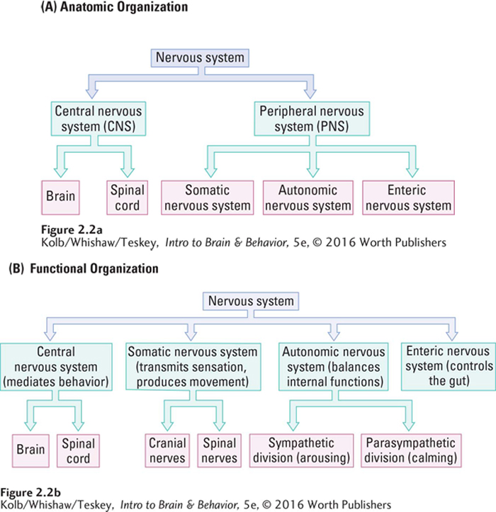
In a functional organization, little changes, but the focus turns to how the parts of the system work together. Neurons in the somatic division of the PNS connect through the cranial and spinal nerves to receptors on the body’s surface and on its muscles. Somatic neurons gather sensory information for the CNS and convey information from the CNS to move muscles of the face, body, and limbs. Similarly, the autonomic division of the PNS enables the CNS to govern the workings of your body’s internal organs—
And so, from a functional standpoint (Figure 2-2B), the major PNS divisions step up to constitute, along with the CNS, an interacting four-
The CNS includes the brain and the spinal cord—
the nervous system core, which mediates behavior. 37
The somatic nervous system (SNS) includes all the spinal and cranial nerves carrying sensory information to the CNS from the muscles, joints, and skin. It also transmits outgoing motor instructions that produce movement.
The autonomic nervous system (ANS) balances the body’s internal organs by producing the rest-
and- digest response through the parasympathetic (calming) nerves or the fight- or- flight response or vigorous activity through the sympathetic (arousing) nerves. The enteric nervous system (ENS), formed by a mesh of neurons embedded in the lining of the gut, controls the gut. The ENS communicates with the CNS via the ANS but mostly operates autonomously.
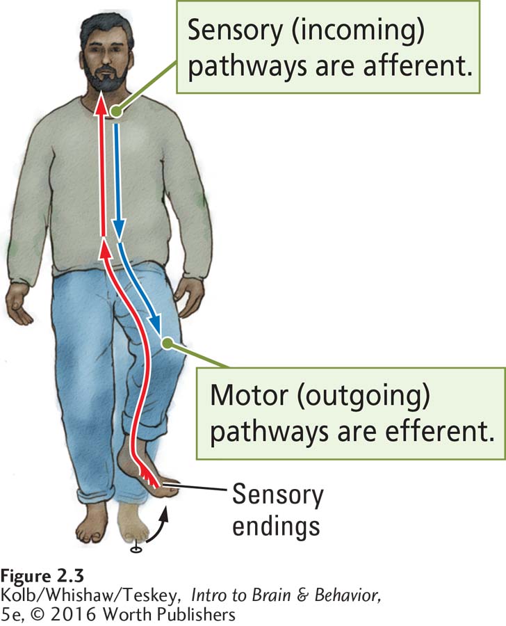
The directional flow of neural information is important. Afferent (incoming) information is sensory, coming into the CNS or one of its parts, whereas efferent (outgoing) information is leaving the CNS or one of its parts. When you step on a tack, the afferent sensory signals are transmitted from the body into the brain and felt as pain. Efferent signals from the brain trigger a motor response: you lift your foot (Figure 2-3).
The Brain’s Surface Features
When buying a new car, people like to look under the hood and examine the engine, the part of the car responsible for most of its behavior—
When it comes to our behavior, the brain is the engine. In many ways, examining a brain for the first time is similar to looking under the car hood. We have a vague sense of what the brain does, but most of us have no sense of how its parts accomplish these tasks. We may not even be able to identify those parts. If you are familiar with the anatomical terms and orientations used in drawings and images of brains, read on. If you prefer to review this terminology before you continue, consult The Basics: Finding Your Way Around the Brain on pages 38–39.
Protecting the Nervous System
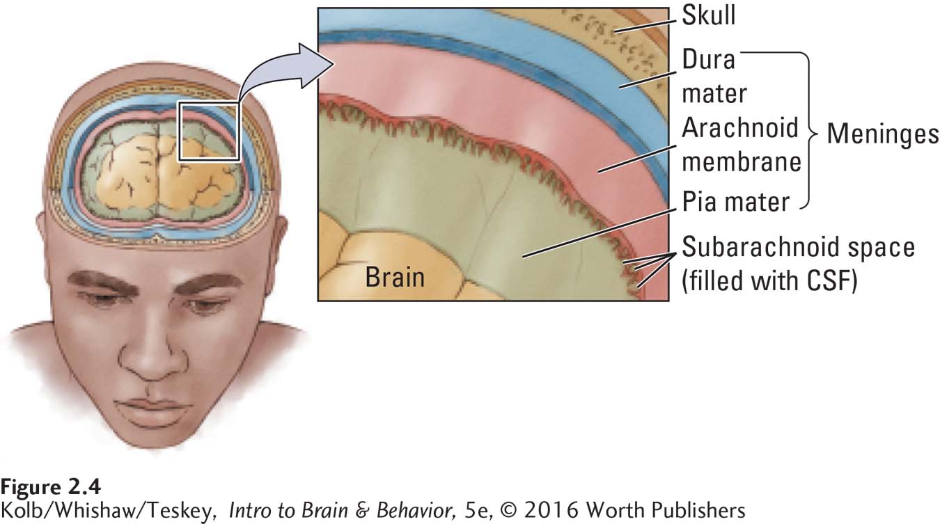
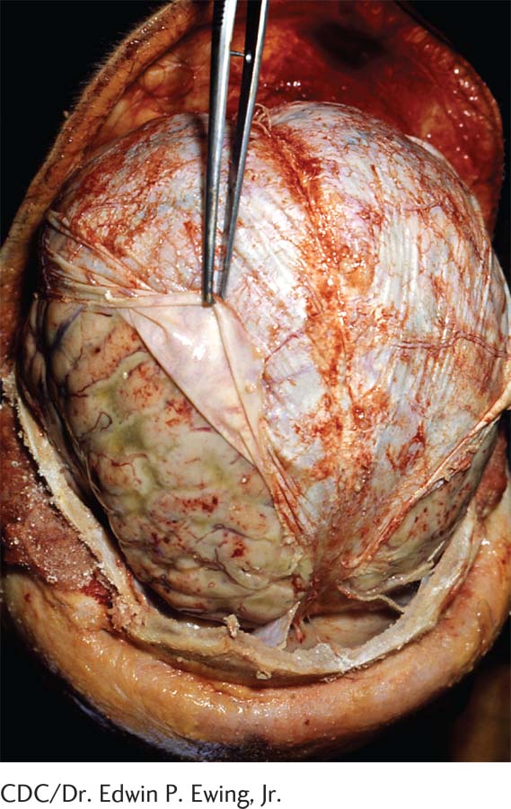
We start our functional overview by opening the hood and observing the brain snug in its home in the skull. The first thing you encounter is not the brain but rather a tough triple-
38
THE BASICS
Finding Your Way Around the Brain
When the first anatomists began to examine the brain with the primitive tools of their time, the names they chose for brain regions often manifested their erroneous assumptions about how the brain works. They named one brain region the gyrus fornicatus because they thought that it had a role in sexual function, but most of this region actually has nothing to do with sexual activity.
A Wonderland of Nomenclature
As time went on, the assumptions and tools of brain research changed, but naming continued to be haphazard and inconsistent. Many brain structures have several names, and terms are often used interchangeably. This peculiar nomenclature arose because research on the brain and behavior spans several centuries and includes scientists of many nationalities and languages.
Early investigators named structures after themselves or objects or ideas. They used various languages, especially Latin, Greek, and English. More recently, investigators have often used numbers or letters, but even this system lacks coherence, because the numbers may be Arabic or Roman and are often used in combination with Greek or Latin letters.
Describing Locations in the Brain
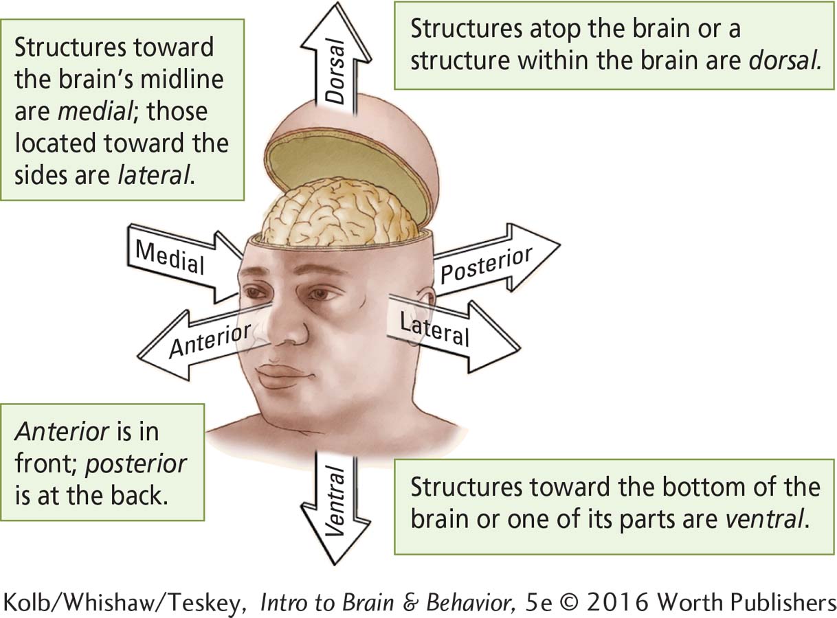
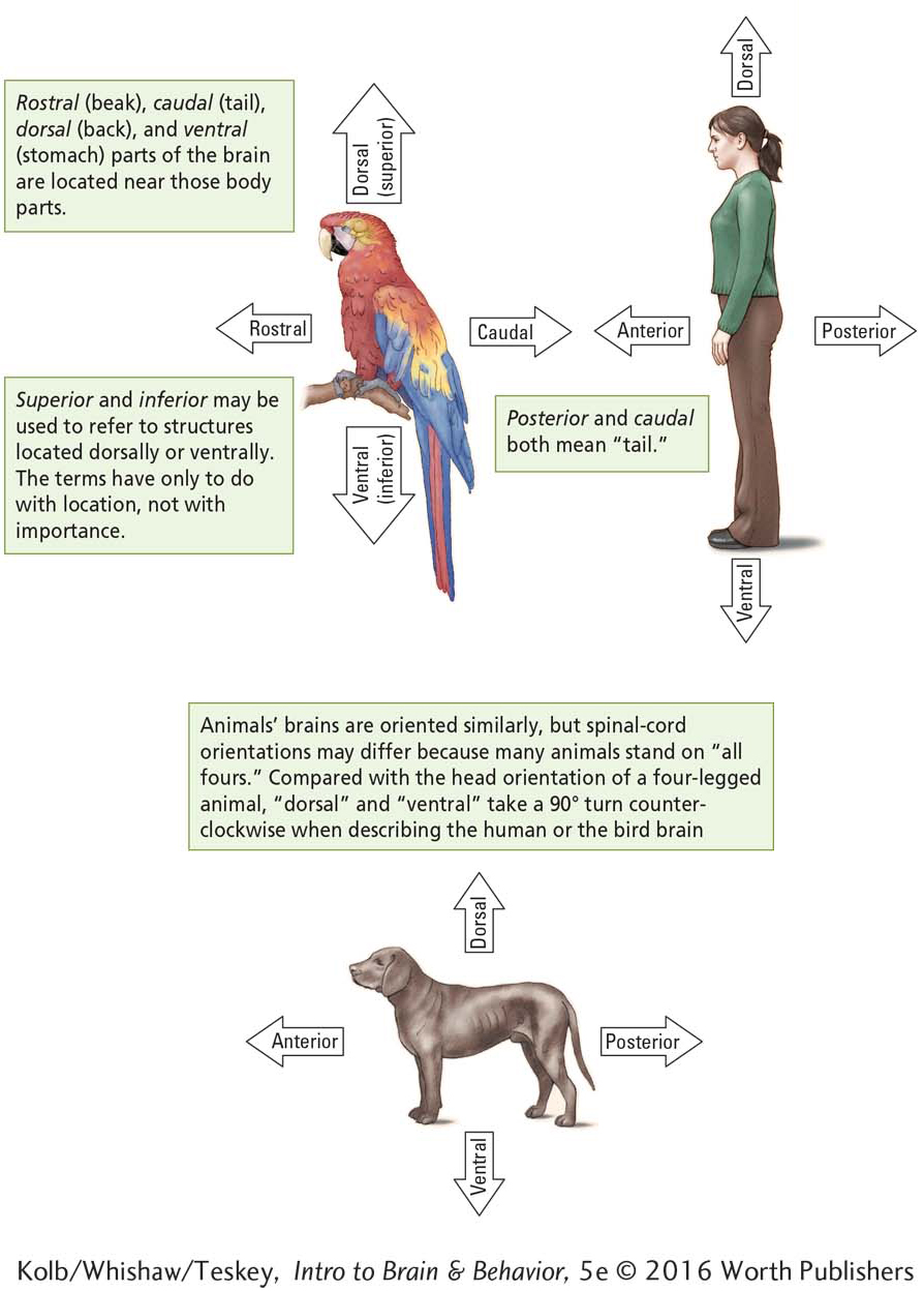
Many names for nervous system structures reflect their anatomical locations with respect to other body parts of the animal, with respect to their relative spatial locations, and with respect to a viewer’s perspective:
Brain–
Body Orientation illustrates brain structure location from the frame of reference of the human face. Spatial Orientation illustrates brain structure location in relation to other body parts and body orientation.
Anatomical Orientation illustrates the direction of a cut, or section, through the human brain (part A) from the perspective of a viewer (part B).
Brain–
Spatial Orientation
39
These orienting terms are derived from Latin. Consult the accompanying Glossary of Anatomical Location and Orientation for easy reference. It is common practice to combine orienting terms. A structure described as dorsolateral, for example, means that it lies up and to the side.

Anatomic Orientation
Finally, the nervous system, like the body, is bilaterally symmetrical: it has a left side and a right side. Structures that lie on the same side are ipsilateral; if they lie on opposite sides, they are contralateral to each other. Structures that occur in each hemisphere are bilateral. Structures that are close to one another are proximal; those far from one another are distal.
Glossary of Anatomical Location and Orientation
| Term | Meaning with respect to the nervous system |
| Anterior | Near or toward the front of the animal or the front of the head (see also frontal and rostral) |
| Caudal | Near or toward the tail of the animal (see also posterior) |
| Coronal | Cut vertically from the crown of the head down; used to reference the plane of a brain section that reveals a frontal view |
| Dorsal | On or toward the back of a four- |
| Frontal | Of the front (see also anterior and rostral); in reference to brain sections, a viewing orientation from the front |
| Horizontal | Cut along the horizon; used to reference the plane of a brain section that reveals a dorsal view |
| Inferior | Below (see also ventral) |
| Lateral | Toward the side of the body or brain |
| Medial | Toward the middle, specifically the body’s midline; in reference to brain sections, a side view of the central structures |
| Posterior | Near or toward the animal’s tail (see also caudal); for human spinal cord, at the back |
| Rostral | Toward the beak (front) of the animal (see also anterior and frontal) |
| Sagittal | Cut lengthways from front to back of the skull to reveal a medial view into the brain from the side; a cut in the midsagittal plane divides the brain into symmetrical halves. |
| Superior | Above (see also dorsal) |
| Ventral | On or toward the belly of four- |
40
Between the arachnoid layer and the pia mater flows cerebrospinal fluid (CSF), a colorless solution of sodium chloride and other salts. CSF cushions the brain so that it can move or expand slightly without pressing on the skull. The symptoms of meningitis, an infection of the meninges and CSF, are described in Clinical Focus 2-2, Meningitis and Encephalitis, on page 42.
Cerebral Geography
After removing the meninges, we can examine the brain’s surface features, most prominently its two nearly symmetrical left and right hemispheres. Figure 2-5 diagrams the left hemisphere of a typical human forebrain oriented in the upright human skull. The outer forebrain consists of a thin, folded film of nerve tissue, the cerebral cortex, detailed in the frontal view in Figure 2-5. The word cortex, Latin for tree bark, is apt, considering the cortex’s heavily folded surface and its location, covering most of the rest of the brain. Unlike the bark on a tree, however, the brain’s folds are not random but rather demarcate its functional cortical zones.
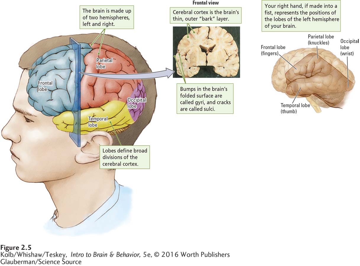
Make a fist with your right hand and hold it up, as shown on the right in Figure 2-5, to represent the positions of the forebrain’s broad divisions, or lobes, in the skull. Each lobe is named for the skull bone it lies beneath.
The forward-
pointing temporal lobe lies at the side of the brain, in approximately the same place as the thumb on your upraised fist. The temporal lobe functions in connection with hearing and with language and musical abilities. Immediately above your thumbnail, your fingers correspond to the location of the frontal lobe, often characterized as performing the brain’s executive functions, such as decision making.
The parietal lobe is at the top of the skull, as represented by your knuckles, behind the frontal lobe and above the temporal lobe. Parietal functions include directing our movements toward a goal or to perform a task, such as grasping an object.
The area at the back of each hemisphere, near your wrist, constitutes the occipital lobe, where visual processing begins.
Examining the Brain’s Surface from All Angles
41
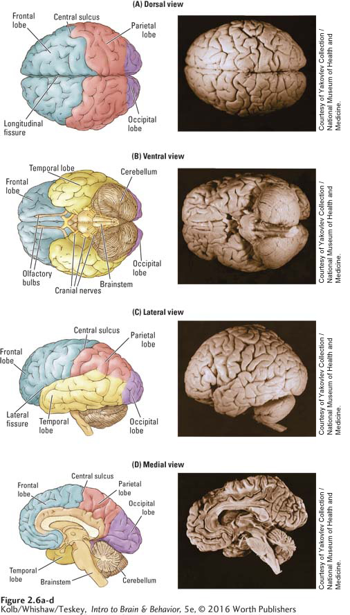
As we look at the dorsal view in Figure 2-6A, the brain’s wrinkled left and right hemispheres resemble a walnut meat taken whole from its shell. These hemispheres constitute the cerebrum, the major forebrain structure and most recently evolved feature of the CNS. Visible from the opposite ventral view in Figure 2-6B are the brainstem, including the wrinkly hemispheres of the smaller cerebellum (Latin for little brain). Both the cerebrum and the brainstem are visible in the lateral and medial views in Figure 2-6C and D.
42
CLINICAL FOCUS 2-2
Meningitis and Encephalitis
Harmful microorganisms can invade the layers of the meninges, particularly the pia mater and the arachnoid layer, as well as the CSF flowing between them, to cause a variety of infections that lead to meningitis. One symptom of this condition, inflammation, places pressure on the brain. Because the space between meninges and skull is slight, unrelieved pressure can lead to delirium and, if the infection progresses, to drowsiness, stupor, and even coma.
Usually the earliest symptom of meningitis is severe headache and a stiff neck (cervical rigidity). Head retraction (tilting the head backward) is an extreme form of cervical rigidity. Convulsions, a common symptom in children, indicate that the inflammation is affecting the brain.
Infection of the brain itself is called encephalitis. Some of its many forms have great historical significance. A century ago, during World War I, a form of encephalitis called sleeping sickness (encephalitis lethargica) reached epidemic proportions. Its first symptom is sleep disturbance. People sleep all day and become wakeful, even excited, at night. Subsequently they show symptoms of Parkinson disease, including severe tremors, muscular rigidity, and difficulty in controlling body movements. Many are completely unable to make voluntary movements, such as walking or even combing their hair. Survivors of sleeping sickness were immortalized by the neurologist Oliver Sacks in the book and movie Awakenings. Sacks died in 2015 at age 82.
Encephalitis symptoms are caused by the death of an area deep in the brain, the substantia nigra (black substance), which you will learn about in Section 2-3. Other forms of encephalitis may have different effects on the brain. For example, Rasmussen encephalitis attacks one cerebral hemisphere in children. In most cases, the only effective treatment is radical: hemispherectomy, surgical removal of the entire affected hemisphere.
Surprisingly, some young children who lose a hemisphere adapt rather well. They may even complete college, literally with half a brain. But intellectual disabilities are a more common outcome of hemispherectomy as a result of encephalitis.
Much of the crinkled-
Looking at the bottom of the brain, the ventral view in Figure 2-6B, we see in the midst of the wrinkled cerebrum and ventral to the cerebellum a smooth, whitish structure with little tubes attached. This central set of structures is the brainstem, the area responsible for most unconscious behavior. The tubes mark out the cranial nerves that run to and from the brain as part of the SNS.
Cerebral Circulation
The brain’s surface appears to be covered with blood vessels. As with the rest of the body, the arteries feed blood to the brain and send it back through veins to the kidneys and lungs for cleaning and oxygenation. The cerebral arteries emerge from the neck to wrap around the outside of the brainstem, cerebrum, and cerebellum, finally penetrating the brain’s surface to nourish its inner regions.
43
Section 16-3 elaborates on the effects of stroke and its treatment.
Three major arteries send blood to the cerebrum—
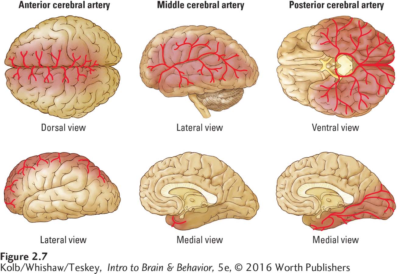
Principle 3: Many brain circuits are crossed.
Because the brain’s connections are crossed, stroke in the left hemisphere affects sensation and movement on the right side of the body. The opposite is true for those with strokes in the right hemisphere. Clinical Focus 2-3, Stroke, on page 45, describes some disruptions that stroke causes, both to the person who has it and to those who care for stroke victims.
The Brain’s Internal Features
The simplest way to examine the inside of something is to cut it in half. Of course, the orientation of the cut affects what we see. Consider slicing through a pear. If we cut from side to side, we cut across the core, providing a dorsal view; if we cut from top to bottom, we cut parallel to the core, providing a medial view. Our impression of the inside of a pear is clearly influenced by how we slice it. The same is true of the brain.
Macro View
We can reveal the brain’s inner features by slicing it parallel to the front of the body, downward through the middle in a coronal section (Figure 2-8A). The resulting frontal view, shown in Figure 2-8B, makes immediately apparent that the brain’s interior is not homogeneous. Both dark and light regions of tissue are visible, and though these regions may not be as distinctive as car engine parts, they nevertheless represent different brain components.
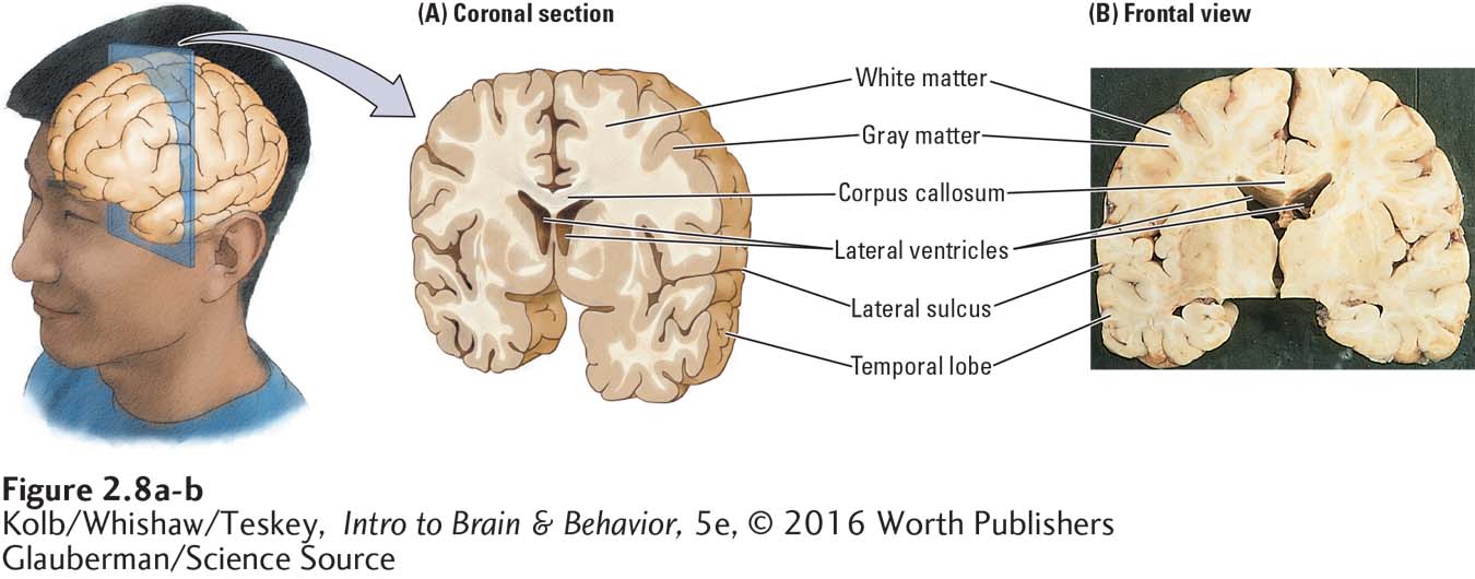
The darker regions are gray matter, largely composed of cell bodies and capillary blood vessels. Gray matter neurons either collect and modify information or support this activity. The lighter regions are white matter, mostly nerve fibers with fatty coverings that produce the white appearance, much as fat droplets in milk make it appear white. White matter fibers form connections between and among the brain’s cells.
44
A second feature, apparent at the center of our frontal view in Figure 2-8B, are the ventricles—two wing-
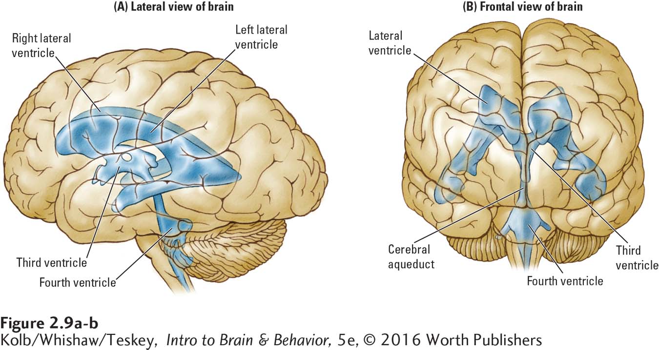
Although the ventricles’ functions are not well understood, researchers think that they play an important role in maintaining brain metabolism. CSF may allow certain compounds access to the brain, and it probably helps the brain excrete metabolic wastes. In the event of trauma to the brain or spinal cord, CSF cushions the blow.
45
CLINICAL FOCUS 2-3
Stroke
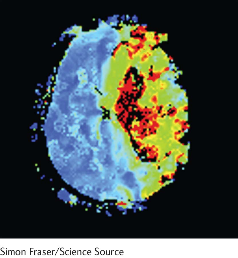
Approximately every minute in the United States, someone has a stroke with obvious visible symptoms—
In addition to visible strokes, at least twice as many silent strokes may occur each year. These ministrokes occur primarily in the white matter and do not produce obvious symptoms. (To view a brief video on silent stroke, go to https:/
Even with the best, fastest medical attention, most stroke patients have some residual motor, sensory, or cognitive deficit. According to the Canadian Stroke Network, for every 10 people who have a stroke, 2 die, 6 are disabled to varying degrees, and 2 recover to a degree but still have a diminished quality of life. Of those who survive, 1 in 10 risk further stroke.
The consequences of stroke are significant for those who have them, their family, and their lifestyle. Consider Mr. Anderson, a 45-
Seven years after his stroke, Mr. Anderson remained unable to speak, but he understood simple conversations. Severe difficulties in moving his right leg required him to use a walker. He could not move the fingers of his right hand and so had difficulty feeding himself, among other tasks. Mr. Anderson will probably never return to his engineering career or drive or get around on his own.
Like him, most stroke survivors require help to perform everyday tasks. Caregivers are often female relatives who give up their own career and other pursuits. Half of the caregivers develop emotional illness, primarily depression or anxiety or both, in a year or so. Lost income and stroke-
We tend to speak of stroke as a single disorder, but two major types of strokes have been identified. In the more common and often less severe ischemic stroke, a blood vessel is blocked, as by a clot. The more severe hemorrhagic stroke results from a burst vessel bleeding into the brain.
The hopeful news is that ischemic stroke can be treated acutely with a drug called tissue plasminogen activator (t-
When patients receive t-
Many people are unable to reach a hospital soon enough for treatment with t-
By taking advantage of developments in neuroimaging, research has shown that it is possible to remove clots from cerebral vessels mechanically (Appireddy et al., 2015). Although quick treatment with t-
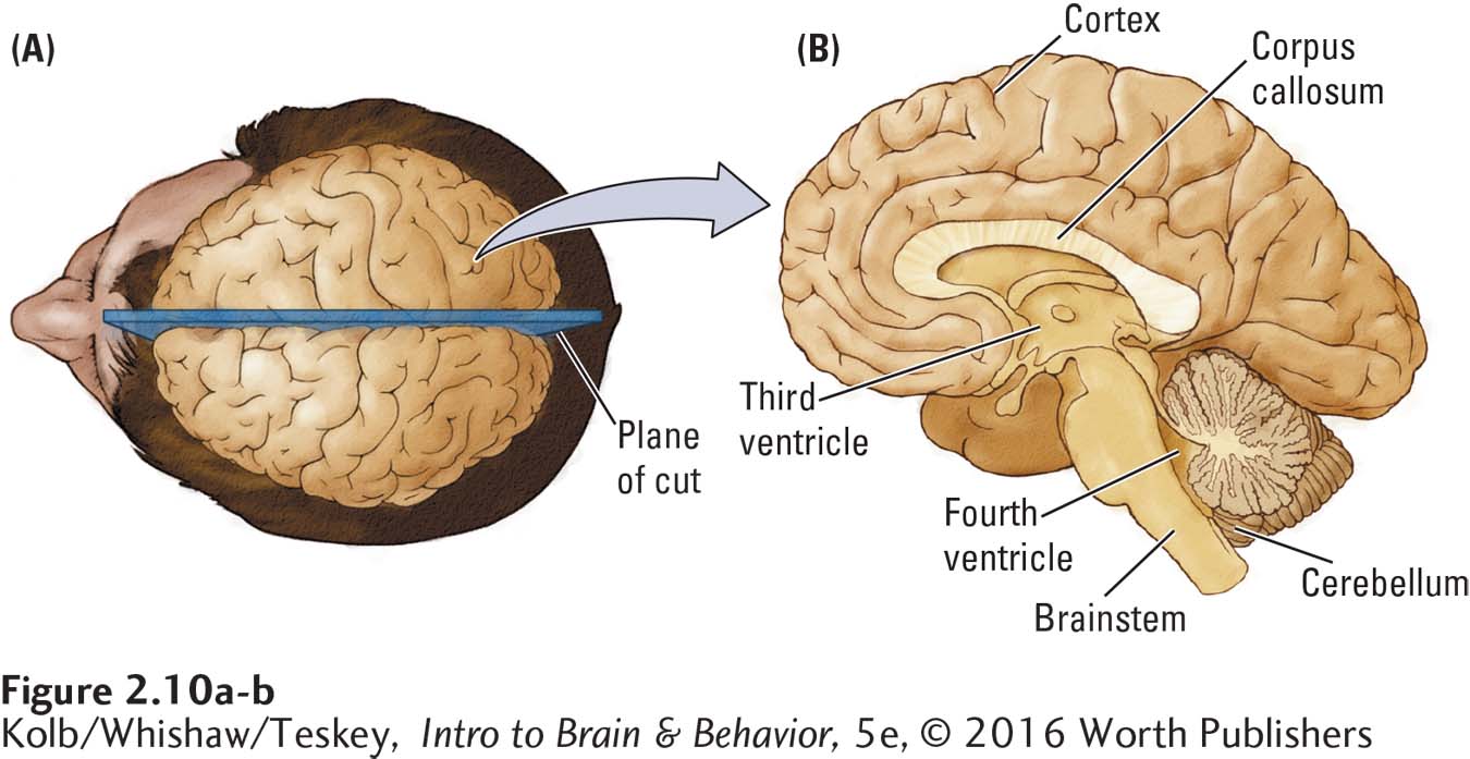
Cutting through the brain vertically from front to back produces a sagittal section (Figure 2-10A). If we make our cut down the brain’s midline, that is, in the midsagittal plane, we divide the cerebrum into its two hemispheres, revealing several distinctive structures in the resulting medial view (Figure 2-10B). One feature is a long band of white matter that runs much of the length of the cerebral hemispheres. This band, the corpus callosum, contains about 200 million nerve fibers that join the two hemispheres and allow them to communicate.
46
Principle 4: The CNS functions on multiple levels.
Figure 2-10B clearly shows that the cortex covers the cerebral hemispheres above the corpus callosum; below it are various internal subcortical regions. The brainstem is a subcortical structure that generally controls basic physiological functions. But many subcortical regions are forebrain structures intimately related to the cortical areas that process motor, sensory, perceptual, and cognitive functions. This relation between the cortex and the subcortex alerts us to the concept that redundant functions exist at many levels of nervous system organization.
Principle 5: The brain is symmetrical and asymmetrical.
If you were to compare medial views of the left and right hemispheres, you would be struck by their symmetry. The brain, in fact, has two of nearly every structure, one on each side. The few one-
Microscopic Inspection: Cells and Fibers
Human brains contain about 86 billion neurons and 87 billion glia. Section 3-1 examines their structures and functions in detail.
The brain’s fundamental units—
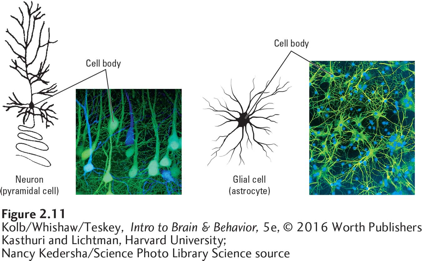
Nancy Kedersha/Science Photo Library Science Source
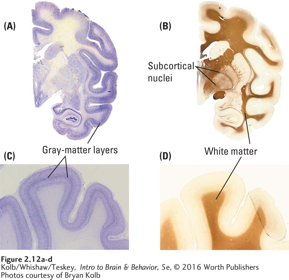
We can see the brain’s internal structures in even greater detail by dyeing their cells with special stains (Figure 2-12). For example, if we use a dye that selectively stains cell bodies, we can see that the neurons in the cortical gray matter lie in layers, revealed by the bands of tissue in Figure 2-12A and C. Each layer contains cells that stain characteristically. Figure 2-12A and B shows that stained subcortical regions are composed of clusters, or nuclei, of similar cells.
47
Although layers and nuclei appear very different, both form functional units in the brain. Whether a particular brain region has layers or nuclei is largely an accident of evolution. By using a stain that selectively dyes neuronal fibers, as shown in Figure 2-12B and D, we can see the borders of the subcortical nuclei more clearly. In addition, we can see that the stained cell bodies lie in regions adjacent to those with most of the fibers.
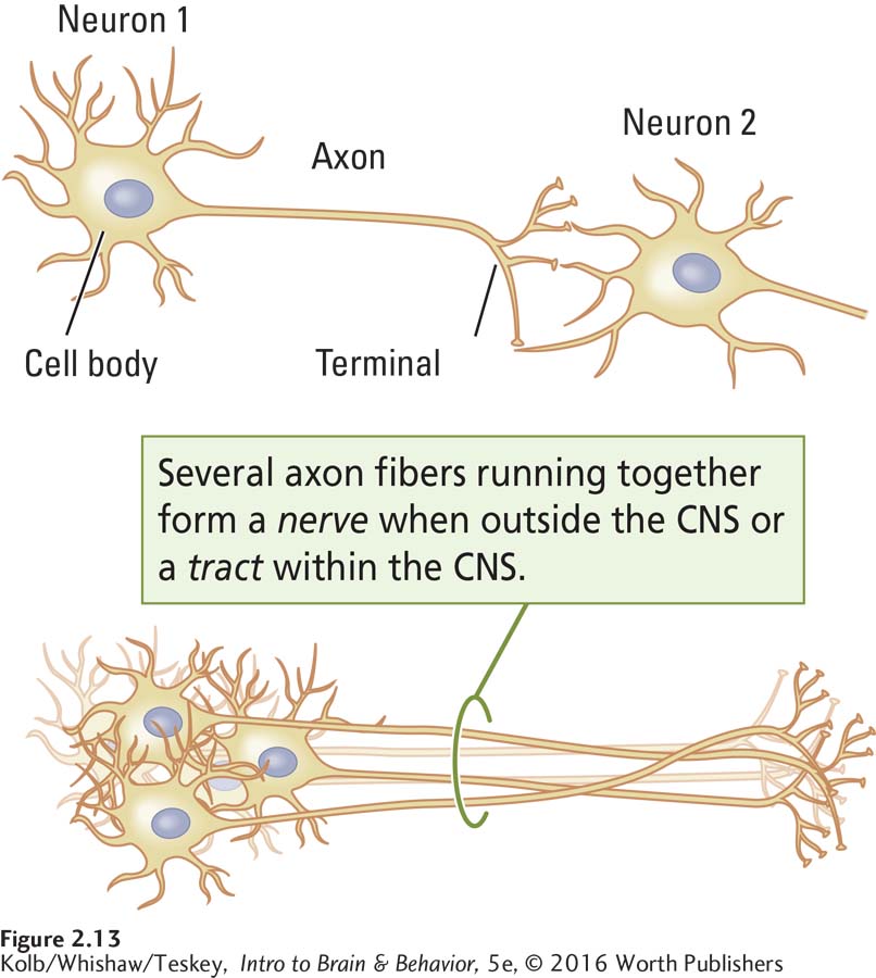
A key feature of neurons is that they are connected to one another by fibers known as axons. When axons run along together, much like the wires that run from a car engine to the dashboard, they form a nerve or tract (Figure 2-13). By convention, a tract is a collection of nerve fibers in the brain and spinal cord, whereas bundles of fibers outside of the CNS are typically called nerves. Thus, the pathway from the eye to the brain is the optic nerve, whereas the pathway from the cerebral cortex to the spinal cord is the corticospinal tract.
2-1 REVIEW
Overview of Brain Function and Structure
Before you continue, check your understanding.
Question 1
The nervous system’s function is to produce movement, or __________, in a perceptual world constructed by the __________.
Question 2
The left and right cerebral hemispheres are each divided into four lobes: __________, __________, __________, and __________.
Question 3
The human nervous system has evolved the potential to change, for example, to adapt to changes in the world or to compensate for injury. This attribute is called __________.
Question 4
Neural tissue is of two main types: (1) __________ forms the connections among cells, and (2) __________ collects and processes incoming (afferent) sensory or outgoing (efferent) information.
Question 5
The nerve fibers that lie in the brain form __________. Outside the brain they are called __________.
Question 6
Chart the human nervous system’s functional organization.
Answers appear in the Self Test section of the book.