2-3 The Central Nervous System: Mediating Behavior
When we look under the hood, we can make some pretty good guesses about what each part of a car engine does. The battery must provide electrical power to run the radio and lights, for example, and because batteries have to be charged, the engine must contain some mechanism for charging them. We can take the same approach to deduce how the parts of the brain function. The part connected to the optic nerve coming from each eye must have something to do with vision. Structures connected to the auditory nerve coming from each ear must have something to do with hearing.
From such simple observations, we can begin to understand how the brain is organized. The real test comes in analyzing actual brain function: how this seeming jumble of parts produces behaviors as complex as human thought. The place to start is the brain’s functional anatomy: learning the name of a particular CNS structure is pointless without also learning something about what it does. We focus now on the names and functions of the three major CNS components: spinal cord, brainstem, and forebrain.
Spinal Cord
Although producing movement is the brain’s principal function, ultimately the spinal cord executes most body movements, usually following instructions from the brain but at times acting independently via the somatic nervous system. To understand how important the spinal cord is, think of the old saying “running around like a chicken with its head cut off.” When a chicken’s head is lopped off for the farmer’s family dinner, the chicken is still capable of running around the barnyard until it collapses from loss of blood. The chicken accomplishes this feat because the spinal cord is acting independently of the brain.
Grasping the spinal cord’s complexity is easier once you realize that it is not a single structure but rather a set of segmented switching stations. As detailed in Section 2-4, each spinal segment receives information from a discrete part of the body and sends out commands to that area. Spinal nerves, which are part of the SNS, carry sensory information to the cord from the skin, muscles, and related structures and in turn send motor instructions to control each muscle.
We explain reflexes in Section 11-4.
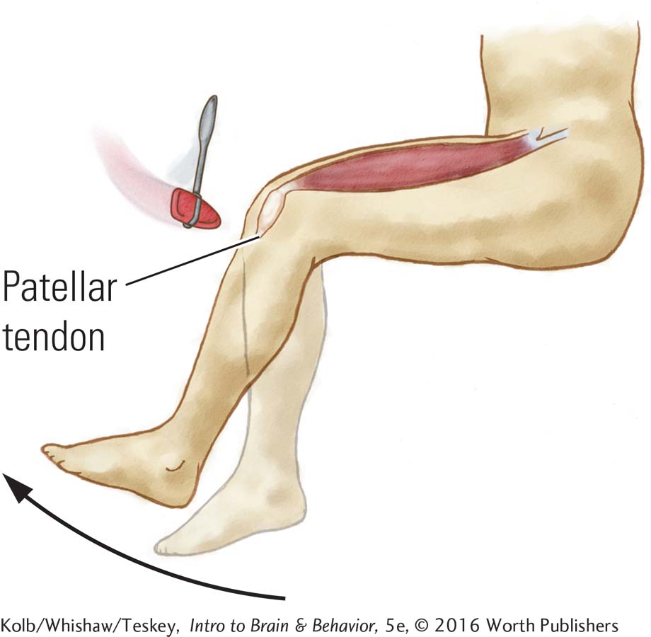
You can demonstrate movement controlled by the spinal cord in your own body by tapping your patellar tendon, just below your kneecap (the patella). The sensory input causes your lower leg to kick out, and try as you might, it is very hard to prevent the movement. Your brain, in other words, has trouble inhibiting this spinal reflex: it is automatic.
Brainstem
51
Alphabetically, afferent comes before efferent: sensory signals must enter the brain before an outgoing signal triggers a motor response.
The brainstem begins where the spinal cord enters the skull and extends upward into the lower areas of the forebrain. The brainstem receives afferent nerves coming in from all of the body’s senses, and it sends efferent nerves out to the spinal cord to control virtually all of the body’s movements except the most complex movements of the fingers and toes. The brainstem, then, both directs movements and creates a sensory world.
In some vertebrates, such as frogs, the entire brain is largely equivalent to the mammalian or avian brainstem. And frogs get along quite well, demonstrating that the brainstem is a fairly sophisticated piece of machinery. If we had only a brainstem, we would still be able to construct a world, but it would be a far simpler sensorimotor world, more like the world a frog experiences.
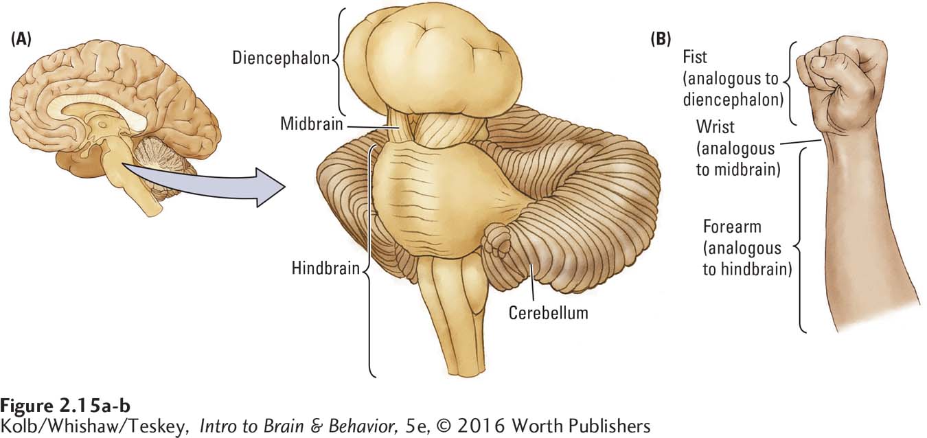
The brainstem, which is responsible for most unconscious behavior, can be divided into three regions: hindbrain, midbrain, and diencephalon, meaning between brain because it borders the brain’s upper and lower parts. In fact, the between-
Principle 7: Sensory and motor divisions permeate the nervous system.
The hindbrain and midbrain are essentially extensions of the spinal cord; they developed first as vertebrate animals evolved a brain at the anterior end of the body. It makes sense, therefore, that these lower brainstem regions should retain a division between structures having sensory functions and those having motor functions, with sensory structures lying dorsal and motor ones ventral, or in upright humans, posterior and anterior.
Each brainstem region performs more than a single task. Each contains various groupings of nuclei that serve various purposes. All three regions, in fact, have both sensory and motor functions. However, the hindbrain is especially important in motor functions, the midbrain in sensory functions, and the diencephalon in integrative sensorimotor tasks. Here we consider the central functions of these three regions; later chapters contain more detailed information about them.
52
Hindbrain
The hindbrain controls motor functions ranging from breathing to balance to fine movements, such as those used in dancing. Its most distinctive structure, and one of the largest in the human brain, is the cerebellum. Its relative size increases with the physical speed and dexterity of a species, as shown in Figure 2-16A.
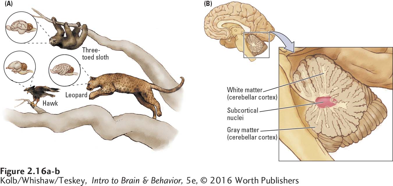
Animals that move relatively slowly (such as a sloth) have a relatively small cerebellum for their body size. Animals that can perform rapid acrobatic movements (such as a hawk or a cat) have a very large cerebellum relative to overall brain size. The human cerebellum, which resembles a cauliflower in the medial view in Figure 2-16B, likewise is important in controlling complex movements. But cerebellar size in humans is also related to cognitive capacity. Relative to other mammals, apes show an expansion of the cerebellum that correlates with increased capacity for planning and executing complex behavioral sequences, including tool use and language (see Barton, 2012).
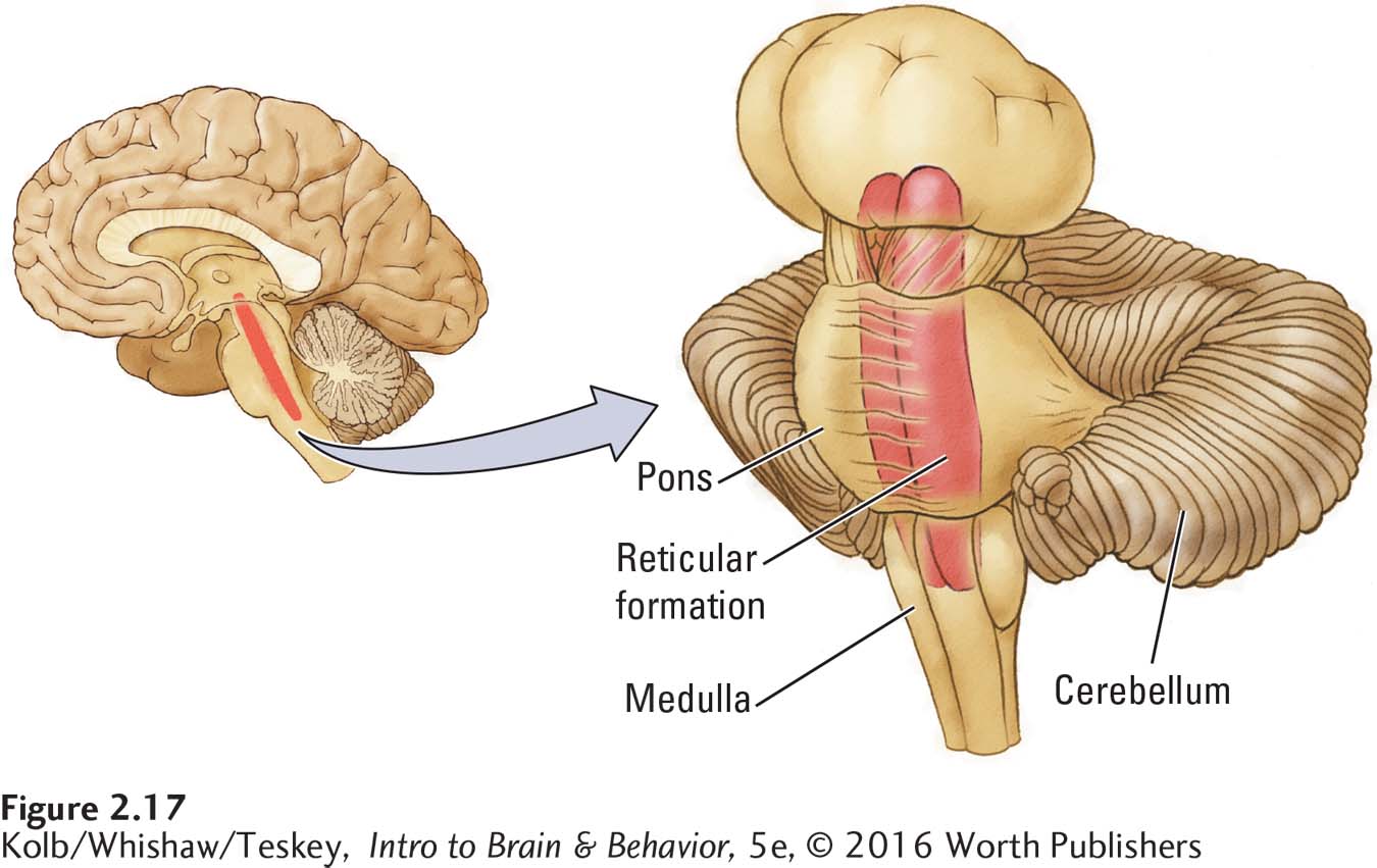
As we look beyond the cerebellum at the rest of the hindbrain, shown in Figure 2-17, we find three subparts: the reticular formation, the pons, and the medulla. Extending the length of the entire brainstem at its core, the reticular formation is a netlike mixture of neurons (gray matter) and nerve fibers (white matter). This nerve net gives the structure the mottled appearance from which its name derives (from Latin rete, meaning net). The reticular formation’s nuclei are localized into small patches along its length. Each has a special function in stimulating the forebrain, such as in waking from sleep.
53
The pons and medulla contain substructures that control many vital body movements. Nuclei in the pons receive inputs from the cerebellum and actually form a bridge from it to the rest of the brain (in Latin, pons means bridge). At the rostral tip of the spinal cord, the medulla’s nuclei regulate such vital functions as breathing and the cardiovascular system. For this reason, a blow to the back of the head can kill you: your breathing stops if the hindbrain control centers are injured.
Midbrain
In the midbrain, a sensory component, the tectum (roof), is dorsal (posterior in upright humans), whereas a motor structure, the tegmentum (floor), is ventral (anterior in humans; Figure 2-18A). The tectum receives a massive amount of sensory information from the eyes and ears. The optic nerve sends a large bundle of fibers to the superior colliculus, whereas the inferior colliculus receives much of its input from auditory pathways. The colliculi function not only to process sensory information but also to produce orienting movements related to sensory inputs, such as turning your head to see a sound’s source.
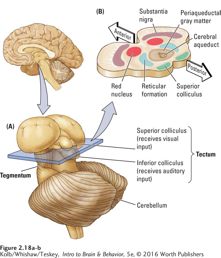
This orienting behavior is not as simple as it may seem. To produce it, the auditory and visual systems must share a map of the external world so that the ears can tell the eyes where to look. If the auditory and visual maps differed, it would be impossible to use the two together. In fact, the colliculi also have a tactile map. After all, if you want to look at what’s making your leg itch, your visual and tactile systems need a common representation of where that place is so you can scratch the itch by moving your arm and hand.
Principle 8: The brain divides sensory input for object recognition and motor control.
Lying ventral to the tectum, the tegmentum (shown in Figure 2-18B in cross section) is composed of many nuclei, largely with movement-
Diencephalon
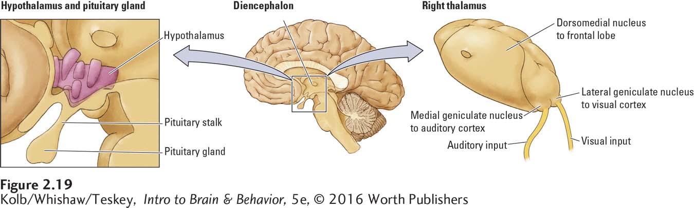
The diencephalon, shown in sagittal section in the center of Figure 2-19, integrates sensory and motor information on its way to the cerebral cortex. Its two principal structures are the hypothalamus and the thalamus. The thalamus—
54
The hypothalamus in each hemisphere lies along the brain’s midline; it is composed of about 22 small nuclei and the nerve fiber systems that pass through it. Its critical function is to control the body’s production of hormones, accomplished via its interactions with the pituitary gland, shown at left in Figure 2-19. Although constituting only about 0.3 percent of the brain’s weight, the hypothalamus takes part in nearly all aspects of behavior, including feeding, sleeping, temperature regulation, sexual and emotional behavior, hormone function, and movement. The hypothalamus is organized and functions more or less similarly across mammals. But sex differences have been found in the structures of some of its parts, owing probably to differences between males and females in activities such as sexual behavior and parenting.
The other principal structure of the diencephalon, the thalamus, is much larger than the hypothalamus, as are its 20-
We examine how thalamic sensory nuclei process incoming information in Sections 9-2, 10-2, 11-4, 12-2, and in Section 14-3, memory pathways.
The routes to the thalamus may be indirect. For example, the route for olfaction traverses several synapses before entering the dorsomedial thalamic nucleus on its way to the forebrain. Analogous sensory regions of the thalamus receive auditory and tactile information, which is subsequently relayed to the respective auditory and tactile cortical regions in each hemisphere. Some thalamic regions have motor functions or, like its dorsomedial nucleus, which connects to most of the frontal lobe, perform integrative tasks.
Forebrain
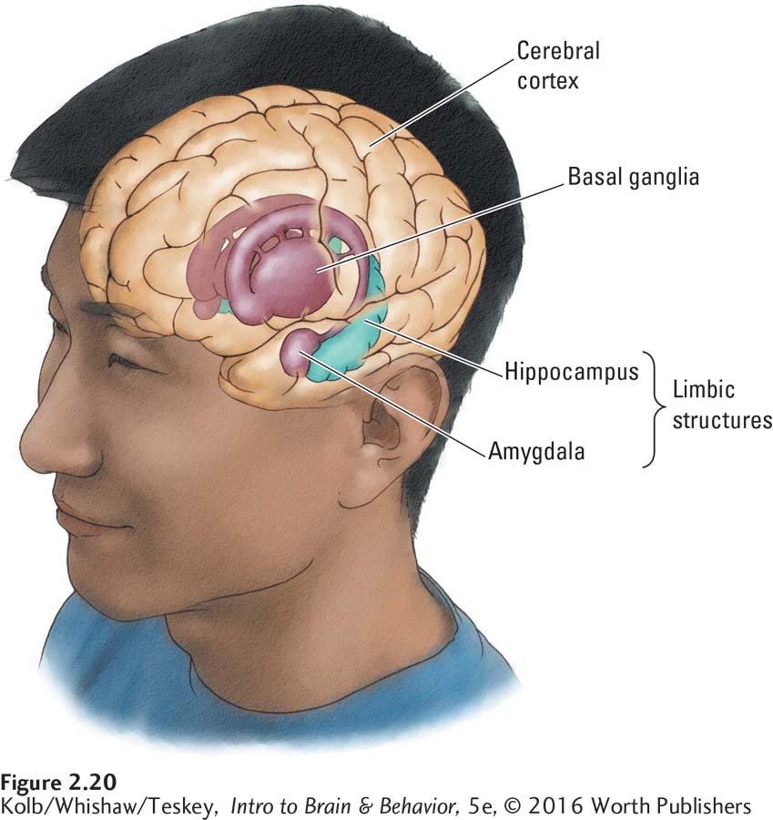
The largest and most recently evolved region of the mammalian brain is the forebrain. Its major internal and external structures are shown in Figure 2-20. Each of its three principal structures has multiple functions. To summarize briefly, the cerebral cortex regulates a host of mental activities ranging from perception to planning; the basal ganglia control voluntary movement; and the limbic system regulates emotions and behaviors that produce and require memory.
Extending our analogy between the brainstem and your forearm, imagine that the fist—
Cerebral Cortex
The forebrain contains two types of cortex, three-
The allocortex plays a role in controlling motivational and emotional states as well as in certain forms of memory. Although neocortex and allocortex have anatomical and functional differences, those distinctions are not critical for most discussions in this book. Therefore, we usually refer to both types of tissue simply as cortex.
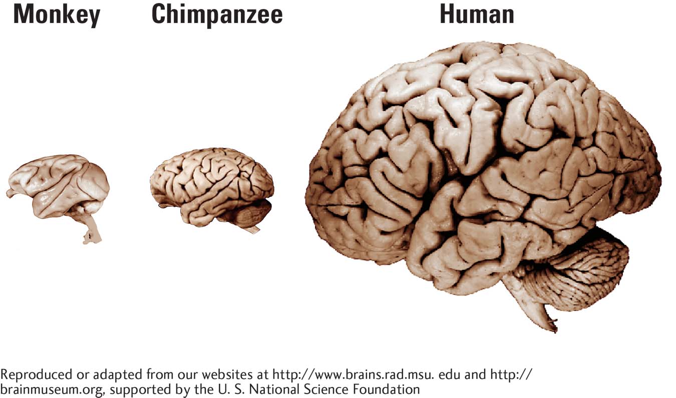
Measured by volume, the cortex makes up most of the forebrain, constituting 80 percent of the human brain overall. It is the brain region that has expanded the most in the course of mammalian evolution. The human neocortex has a surface area as large as 2500 square centimeters but a thickness of only 1.5 to 3.0 millimeters, an area equivalent to about four pages of this book. By contrast, a chimpanzee has a cortical area equivalent to about one page.
55
The pattern of sulci and gyri formed by the folding of the cortex varies across species. Some, such as rats, have no sulci or gyri, whereas in carnivores, such as cats, the gyri form a longitudinal pattern. In primates, the sulci and gyri form a more diffuse pattern.
Cortical Lobes
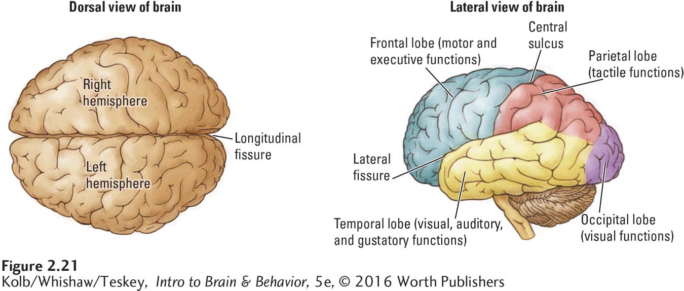
To review, the human cortex consists of the nearly symmetrical left and right hemispheres, which are separated by the longitudinal fissure, shown at left in Figure 2-21. As shown at right, each hemisphere is subdivided into four lobes corresponding to the skull bones overlying them: frontal, temporal, parietal, and occipital. Unfortunately, bone location and brain function are unrelated. As a result, the cortical lobes are rather arbitrarily defined anatomical regions that include many functional zones.
Principle 9: Brain functions are localized and distributed.
Nevertheless, we can attach some gross functions to each lobe. The three posterior lobes have sensory functions: the occipital lobe is visual; the parietal lobe is tactile; and the temporal lobe is visual, auditory, and gustatory. In contrast, the frontal lobe is motor and is sometimes called the brain’s executive because it integrates sensory and motor functions and formulates plans of action. We can also predict some effects of injuries to each lobe:
People with an injured occipital lobe have deficits in processing visual information. Although they may perceive light versus dark, they may be unable to identify either the shape or the color of objects.
Injuries to the parietal lobe make it difficult to identify or locate stimulation on the skin. Deficits in moving the arms and hands to points in space may occur.
Temporal lobe injuries result in difficulty recognizing sounds, although unlike people with occipital injuries, those with temporal injury can still recognize that they are hearing something. Temporal lobe injuries can also produce difficulties in processing complex visual information, such as faces.
Individuals with frontal lobe injuries may have difficulties organizing and evaluating their ongoing behavior as well as planning for the future.
Anatomical features presented in Section 9-2 define occipital lobe boundaries.
Fissures and sulci often establish the boundaries of cortical lobes (Figure 2-21, right). For instance, in humans, the central sulcus and lateral fissure form the boundaries of each frontal lobe as well as the boundaries of each parietal lobe lying posterior to the central sulcus. The lateral fissure demarcates each temporal lobe, forming its dorsal boundary. The occipital lobes are not so clearly separated from the parietal and temporal lobes because no large fissure marks their boundaries.
Cortical Layers
56
In the neocortex, six layers of gray matter sit atop a layer of white matter. The allocortex, by contrast, has three or four layers of gray matter. The six layers of the neocortex have distinct characteristics:
Different layers have different types of cells.
The cell density varies from layer to layer, ranging from virtually no cells in layer I (the top layer) to very dense cell packing in layer IV of the neocortex (Figure 2-22).
Other differences in appearance are both regional and functional.
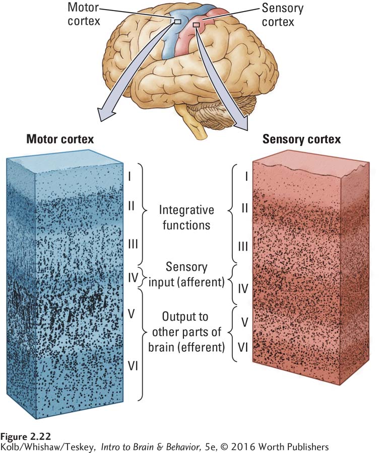
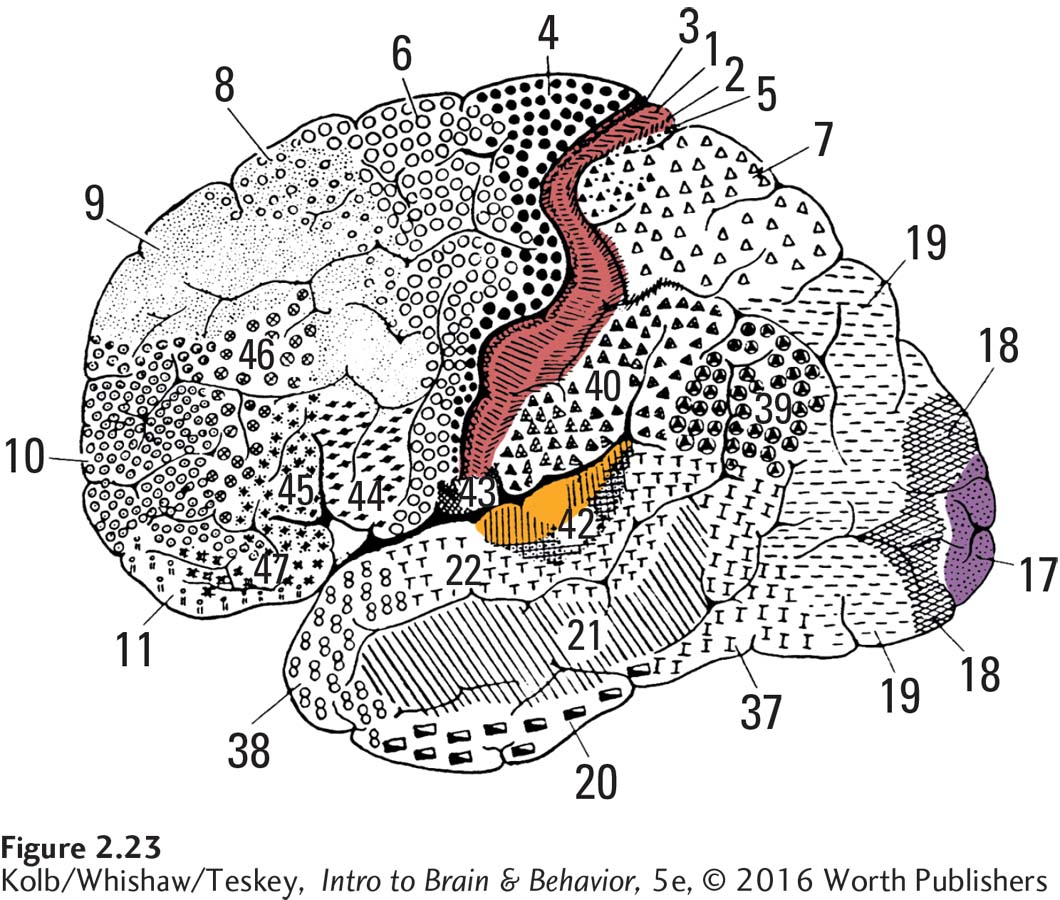
These visible differences led neuroanatomists to map the cortex a century ago. Korbinian Brodmann developed the map in Figure 2-23 around 1905. Because these maps are based on cell characteristics, the subject of cytology, they are called cytoarchitectonic maps. For example, viewed through a microscope, sensory cortex in the parietal lobe, shown in red in Figure 2-22, has a distinct layer IV. Motor cortex in the frontal lobe, shown in blue in Figure 2-22, has a distinctive layer V. Layer IV is afferent; layer V is efferent. It makes sense that a sensory region has a large input layer, whereas a motor region has a large output layer.
Staining cortical tissue can reveal chemical differences between cells and layers. Some regions are rich in one chemical, others rich in another. These differences presumably relate to functional specialization of cortical areas.
The one significant difference between the organization of the cortex and the organization of other brain parts is its range of connections. Unlike most structures, which connect only to certain brain regions, the cortex is connected to virtually all other parts of the brain. The cortex, in other words, is the ultimate meddler. It takes part in everything. This fact not only makes it difficult to identify specific cortical functions but also complicates our study of the rest of the brain, because we must always consider the cortex’s role in other brain regions.
Consider your perception of clouds. You have no doubt gazed up at clouds on a summer day and imagined sailing ships, elephants, faces, and countless other objects. Although a cloud does not really look like an elephant, you can concoct an image of one if you impose your frontal cortex—
The cortex influences many behaviors besides object perception. It influences our cravings for foods, our lust for things (or people), and how we interpret the meaning of abstract concepts, words, and images. The cortex ultimately creates our reality, and one reason it serves this function is that it is so well-
57
Basal Ganglia
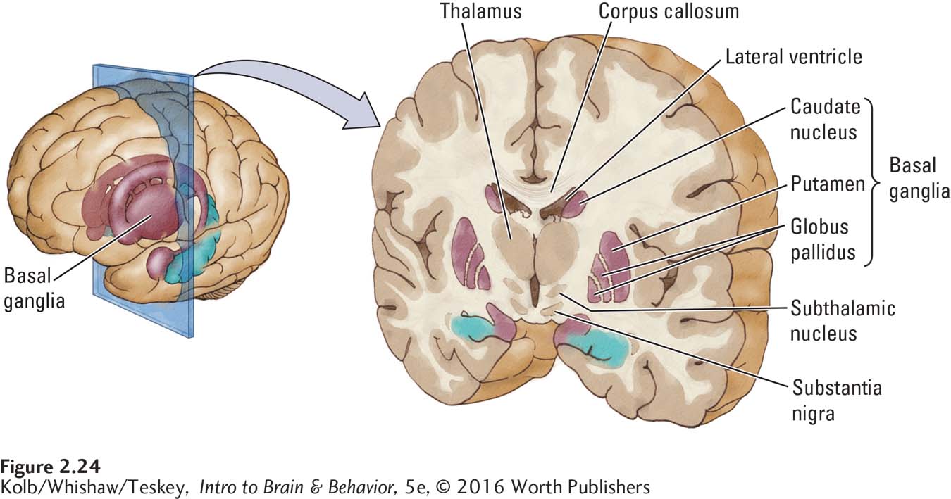
A collection of nuclei that lie in the forebrain just below the white matter of the cortex, the basal ganglia consist of three principal structures: the caudate nucleus, the putamen, and the globus pallidus, all shown in Figure 2-24. Together with the thalamus and two closely associated nuclei, the substantia nigra and subthalamic nucleus, the basal ganglia form a system that functions primarily to control voluntary movement.
Details on Parkinson disease appear in Focus boxes 5-2, 5-3, and 5-4, Sections 11-3 and 16-3. Focus 11-4 details Tourette syndrome.
We can observe the functions of the basal ganglia by analyzing the behavior resulting from the many diseases that interfere with their healthy functioning. Parkinson disease, a motor system disorder characterized by severe tremors, muscular rigidity, and a reduction in voluntary movement, is among the most common movement disorders among the elderly. People with Parkinsonism take short, shuffling steps; display bent posture; and may need a walker to get around. Many have almost continuous hand tremors and sometimes head tremors as well. Another disorder of the basal ganglia is Tourette syndrome, characterized by various motor tics; involuntary vocalizations (including curse words and animal sounds); and odd, involuntary body movements, especially of the face and head.
Neither Parkinsonism nor Tourette syndrome is a disorder of producing movements, as in paralysis. Rather, they are disorders of controlling movements. The basal ganglia, therefore, must play a critical role in controlling and coordinating movement patterns rather than in activating the muscles to move.
Limbic System
In the 1930s, psychiatry was dominated by the theories of Sigmund Freud, who emphasized the roles of sexuality and emotion in human behavior. At the time, the brain regions controlling these behaviors had not been identified, and coincidentally, a group of brain structures collectively called the limbic lobe had no known function. It was a simple step to thinking that perhaps the limbic structures played a central role in sexuality and emotion.
One sign that this hypothesis might be correct came from James Papez (1937), who discovered that people with rabies have infection in the limbic structures, and one symptom of rabies is heightened emotionality. We now know that such a simple view is inaccurate. In fact, the limbic system is not a unitary system at all. And although some limbic structures have roles in emotional and sexual behaviors, limbic structures serve other functions too, including contributing to memory and motivation.
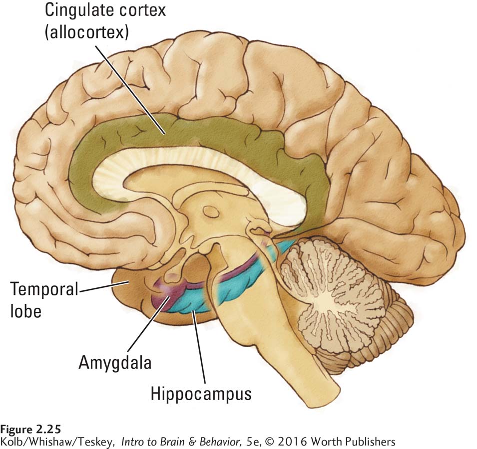
Figure 2-25 diagrams the principal limbic structures Papez proposed. The hippocampus, cingulate cortex (a type of allocortex), and associated structures participate in certain memory functions as well as in controlling navigation in space. Many limbic structures, in particular the amygdala, are also believed to contribute to the rewarding properties of psychoactive drugs and other potentially addictive substances and behaviors. Repeated exposure to drugs such as amphetamine or nicotine produces both chemical and structural changes in the cingulate cortex and hippocampus, among other structures.
58

Removal of the amygdala produces truly startling changes in emotional behavior. A cat with the amygdala removed will wander through a colony of monkeys, completely undisturbed by their hooting and threats. No self-
Olfactory System
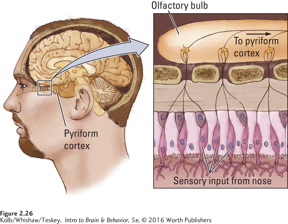
At the very front of the brain lie the olfactory bulbs, the organs responsible for our sense of smell. The olfactory system is unique among human senses, as Figure 2-26 shows, because it is almost entirely a forebrain structure. The other sensory systems project most of their inputs from the sensory receptors to the midbrain and thalamus. Olfactory input takes a less direct route: the olfactory bulb sends most of its inputs to a specialized region, the pyriform cortex, on the brain’s ventral surface. From there, sensory input progresses to the amygdala and the dorsomedial thalamus (see Figure 2-19, right), which routes it to the frontal cortex.
Smell is one of the first senses to have evolved in animals, yet curiously, the olfactory system lies at the front of the human brain and is considered part of the forebrain (see the ventral view in Figure 2-6). This is partly an accident of evolution. The olfactory bulbs lie near the olfactory receptors in the nasal cavity. Although they send their inputs to the pyriform cortex in mammals, their input to the brainstem is more direct in simpler brains.
Section 12-2 considers the chemical senses smell and taste in the context of emotional and motivated behavior.
Compared with the olfactory bulbs of animals such as rats, cats, and dogs, which depend more heavily on smell than we do, the human olfactory bulb is relatively small. Nonetheless, it is very sensitive, allowing us to distinguish a surprisingly large number of odors. Smell plays important roles in various aspects of our feeding and sexual behavior.
2-3 REVIEW
The Central Nervous System: Mediating Behavior
Before you continue, check your understanding.
Question 1
The three functionally distinct sections of the CNS—
Question 2
The __________ can perceive sensations from the skin and muscles and produce movements independent of the brain.
Question 3
The brainstem includes three functional regions. The __________ is an extension of the spinal cord; the __________ is the first brain region to receive sensory inputs; and the __________ integrates sensory and motor information on its way to the cerebral cortex.
59
Question 4
The __________ coordinates fine motor movements and various cognitive functions.
Question 5
The forebrain’s subcortical regions include the __________, which control voluntary movement, and the __________, which controls mood, motivation, and some forms of memory.
Question 6
The two types of cerebral cortex are the older __________ and the __________, which features layers that vary in density to perform __________, __________, and functions.
Question 7
Briefly describe the functions performed by the forebrain.
Answers appear in the Self Test section of the book.