3-1 Cells of the Nervous System
Cells have been likened to factories in that they make a product, proteins, the building blocks of our bodies. But cells are more than factories. Cells use the proteins they produce to participate in orchestrating our behavior.
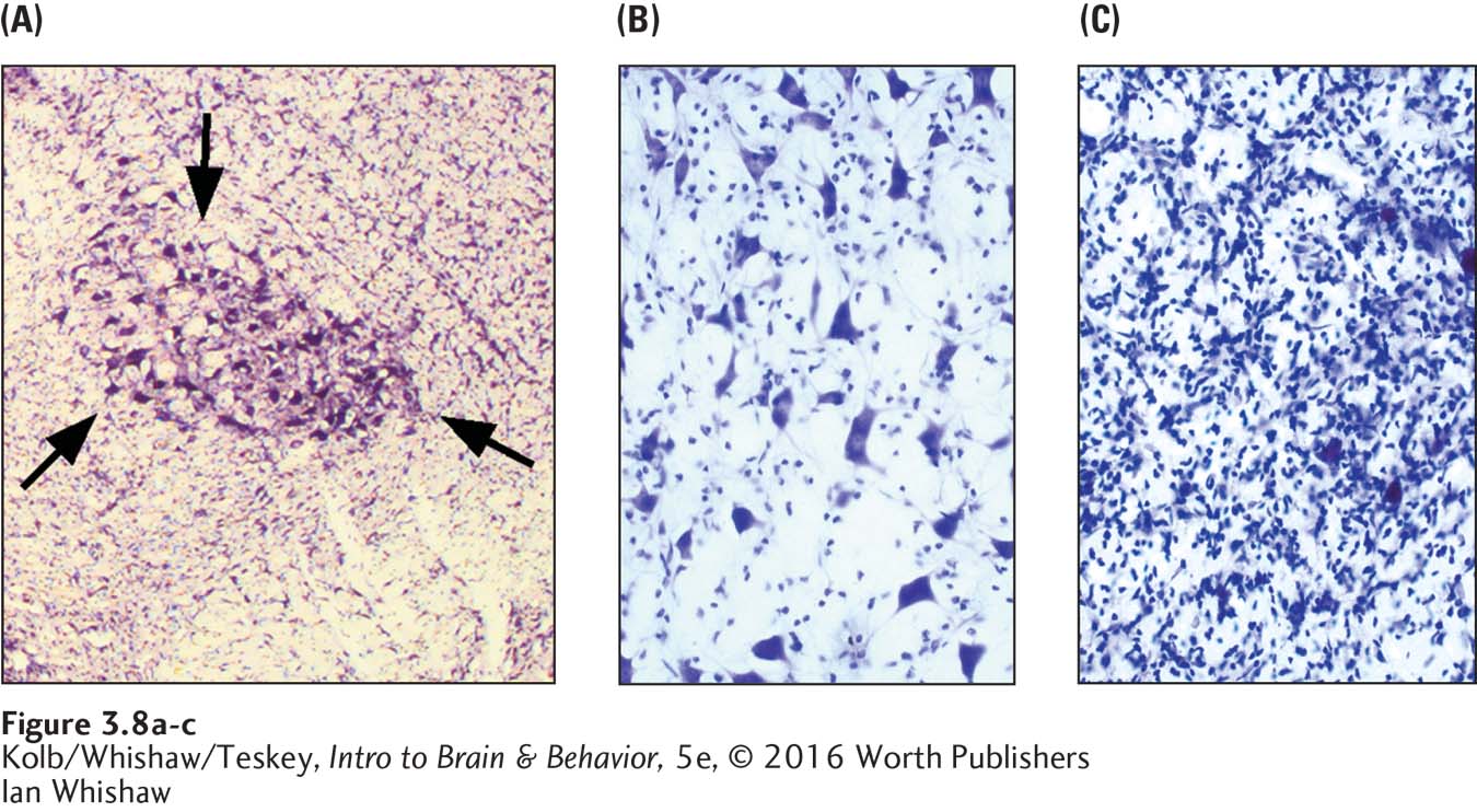
Nervous system cells are small, are packed tightly together, and have the consistency of jelly. To see a brain cell, the observer must distinguish it from surrounding cells, magnified under a microscope. The first anatomists to study brain cells developed methods of highlighting individual cells in nervous system tissue. To make the tissue firm, they soaked it in formaldehyde to preserve and fix the tissue by binding together its constituent protein molecules. The fixed tissue was then sliced in thin sheets (sectioned), colored (stained) with various dyes, and placed under a microscope for viewing.
75
RESEARCH FOCUS 3-2
Brainbow: Rainbow Neurons
Were it not for the discovery of stains that can highlight brain cell features, their complexity and connections would remain unknown. Jean Livet (2007) and his colleagues at Harvard University developed a transgenic technique that labels different neurons by highlighting them with distinct colors, a technique called Brainbow, a play on the word rainbow. (Transgenic techniques are a form of genetic engineering discussed in Section 3-3.)
In the same way a television monitor produces the full range of colors that the human eye can see by mixing only red, green, and blue, the Brainbow scientists introduced genes that produce cyan (blue), green, and red fluorescent proteins into mice cells. The red gene is obtained from coral, and the blue and green genes are obtained from jellyfish. (The 2008 Nobel Prize in chemistry was awarded to Roger Tsien, Osamu Shimomura, and Martin Chalfie for their discovery and development of fluorescent proteins in coral and jellyfish.)
The mice also received a bacterial gene called Cre. Cre activates the color genes inside each cell, but owing to chance factors, the extent to which each gene is activated varies. As the mice develop, the variable expression of the color-
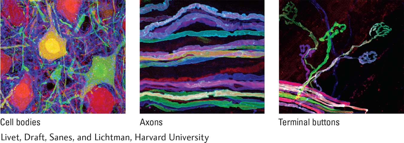
Because individual cells can be visualized, Brainbow offers a way to describe where each neuron sends its processes and how it interconnects with other neurons. You have probably seen an electrical power cord in which the different wires have different colors (black, white, red) that signify what they do and how they should be connected. By visualizing living brain tissue in a dish, Brainbow provides a method for examining changes in neural circuits with the passage of time.
In the future, Brainbow will prove useful for examining populations of cells and their connections—
Scientists continue to fix, slice, and stain brain tissue and to improve on ways of visualizing cells to provide insights into what cells do. Visualization is aided not only by using dyes that either color an individual cell completely; color some cellular components. such as its proteins; or as described in Research Focus 3-2, Brainbow: Rainbow Neurons, color the cell only when it is engaged in a particular activity.
Scientists have developed other techniques of viewing living cells in the nervous system or viewing cells that are cultured in a dish with nurturing fluids. In doing so, they use staining techniques to produce an image of the cell and to allow its activity to be viewed and controlled. Investigators even implant tiny microscopes in the brain to view the activity of its neurons (Chen et al., 2013). There remains, however, the problem of making sense of what you see. Different brain samples can yield different images, and different people can interpret the images in different ways.
So began a controversy between two great scientists—
76
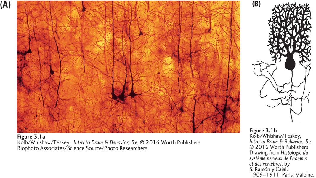
(B) Drawing from Histologie du système nerveux de l’homme et des vertèbres, by S. Ramón y Cajal, 1909–
Golgi never revealed just how he came to develop his staining technique.
The image is beautiful and intriguing, but what do you make of it? To Golgi, this structure suggested that the nervous system is an interconnected network of fibers. He thought that information flowed around this “nerve net,” like water running through pipes, and so produced behavior. His theory was reasonable, given what he saw.
But Santiago Ramón y Cajal came to a different conclusion. He used Golgi’s stain to study chick embryos’ brain tissue. He assumed that their developing nervous system would be simpler and easier to understand than an adult’s. Figure 3-1B shows an image he rendered from the neural cells using the Golgi stain. Cajal concluded that the nervous system is made up of such discrete cells, which begin life as a rather simple structure that becomes more complex with age. When mature, each cell consists of a main body with many extensions projecting from it.
The structure looks something like a plant, with branches coming out of the top and roots coming out of the bottom. Cajal showed that neurons come in many shapes and sizes. Cajal’s neuron theory—that neurons are the nervous system’s functional units—
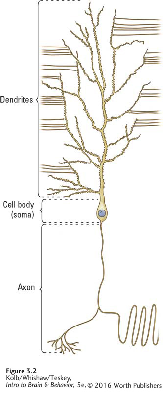
Figure 3-2 shows the three basic subdivisions of a neuron. The core region is called the cell body, or soma (Greek, meaning body; the root of words such as somatic). A neuron’s branching extensions, or dendrites (from Greek for tree), collect information from other cells, and its main root is the single axon (Greek for axle), which carries messages to other neurons. A neuron has only one axon, but most have many dendrites. Some neurons have so many dendrites that they look like a garden hedge, as Cajal’s drawing in Figure 3-1B illustrates.
The human nervous system contains 86 billion neurons (Herculano-
Neurons: The Basis of Information Processing
As the information-
77
Some scientists think that a specific function can be assigned to an individual neuron. For example, many birds learn to sing during the breeding season, and this learning is associated with the seasonal development of new neurons (Bertram et al., 2014). To produce most behaviors in most species, however, scientists think that neurons work together in groups of many hundreds to many thousands. It is important, then, to understand not only how neurons function but also how they interconnect and influence one another.
Functional groups of neurons, or neural networks, connect wide areas of the brain and spinal cord. The loss of a neuron or two from a network is no more noticeable than the loss of one or two voices from a cheering crowd. It is the crowd that produces the overall sound, not each person. In much the same way, although neuroscientists call neurons the information-
Scientists also speak informally about a particular neuron’s structure as if that structure never changes. But neurons are the essence of plasticity. If you view fresh brain tissue through a microscope, the neurons reveal themselves to be surprisingly active, producing new branches, losing old ones, and making and losing connections with each other as you watch. This dynamic activity underlies both the constancies and the changes in our behavior.
Another important property of most neurons is their longevity. At a few locations in the human nervous system, ongoing production of new neurons does take place throughout life, and some behavior does depend on the production of new neurons. But most of our CNS neurons are with us for life. Although they change, they are not replaced when lost. For this reason, if the brain or spinal cord is damaged, functional recovery is often poor. In some nonhuman animal species, neurons can be replaced. The variability in neuronal longevity and replacement are important properties to scientists who study diseases of aging, disorders associated with age-
Structure and Function of the Neuron
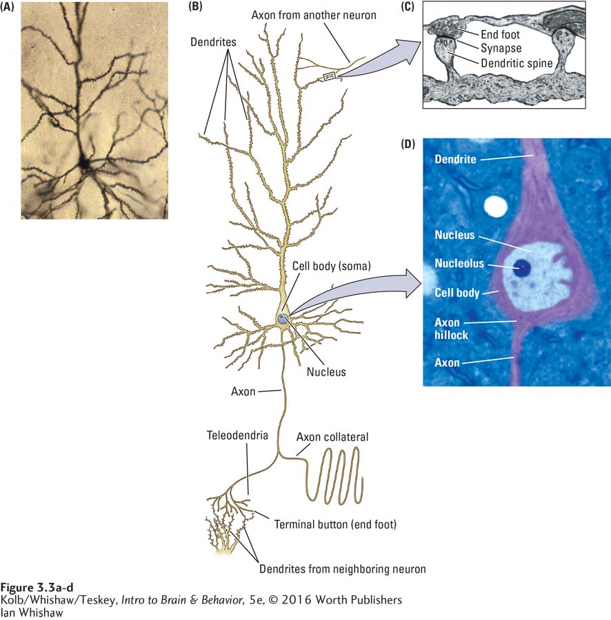
Figure 3-3 on page 78 details the external and internal features common to neurons. The cell’s surface area is increased immensely by its extensions into dendrites and an axon (Figure 3-3A and B). The dendritic area is further increased by many small protrusions called dendritic spines (Figure 3-3C). A neuron may have up to 20 dendrites, each may have one to many branches, and the spines on the branches may number in the thousands. Dendrites collect information from other cells, and the spines are the points of contact with those neurons. The extent of a cell’s branches and its spine number correspond to its information-
Each neuron has but a single axon to carry messages to other neurons. The axon begins at one end of the cell body at an expansion known as the axon hillock (little hill), shown in Figure 3-3D. The axon may branch out into one or many axon collaterals, which usually emerge from it at right angles, as shown at the bottom of Figure 3-3B.
The axon’s lower tip may divide into multiple smaller branches (telodendria, or end branches). At the end of each telodendrion is a knob called an end foot, or terminal button. The terminal button sits very close to but usually does not touch a dendritic spine or some other part of another cell (Figure 3-3C). This near-
78
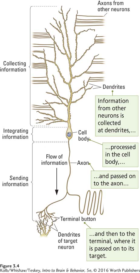
Chapter 4 describes how neurons transmit information; here, we simply generalize about neuronal function by examining shape. Imagine looking down on a river system from an airplane. You see many small streams merging to make creeks, which join to form tributaries, which join to form the main river channel. As the river reaches its delta, it breaks up into many smaller channels again before discharging its contents into the sea.
The general shape of a neuron suggests that it works in a broadly similar way to a river. As illustrated in Figure 3-4, the neuron collects information from many sources on its dendrites. It channels the information to its axon, which can send out only a single message. The eventual branching of the axon then allows the message to be sent to many targets.
Three Functions of Neurons
Neurons of varying shapes and sizes are structured to perform specialized functions. Sensory neurons (Figure 3-5A) conduct information from the sensory receptors in or on the body into the spinal cord and brain. Interneurons (Figure 3-5B) associate sensory and motor activity in the CNS, and motor neurons (Figure 3-5C) carry information from the brain and spinal cord out to the body’s muscles.
79
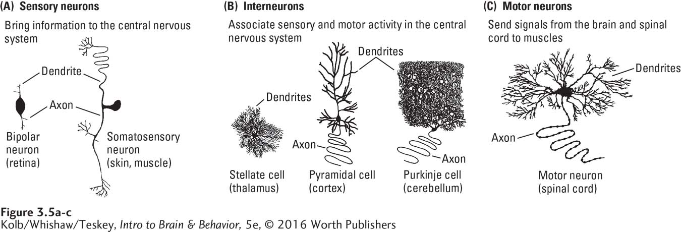
SENSORY NEURONS Sensory neurons are the simplest structurally. A bipolar neuron found in the retina of the eye, for example, has a single short dendrite on one side of its cell body and a single short axon on the other side. Bipolar neurons transmit afferent (incoming) sensory information from the retina’s light receptors to the neurons that carry information into the brain’s visual centers.
A bit more structurally complicated is the somatosensory neuron, which brings sensory information from the body into the spinal cord, a long distance. Structurally, the somatosensory dendrite connects directly to its axon, so the cell body sits to one side of this long pathway.
INTERNEURONS Also called association cells because they link up sensory and motor neurons, interneurons branch extensively, the better to collect information from many sources. Animals with large brains have more interneurons, and their interneurons have more branches, than those of small-
A pyramidal cell has a long axon, a pyramid-
MOTOR NEURONS To collect information from many sources, motor neurons have extensive dendritic networks, large cell bodies, and long axons that connect to muscles. Motor neurons reside in the lower brainstem and spinal cord. All efferent (outgoing) neural information must pass through them to reach the muscles.
Neuronal Networks
Sensory neurons collect afferent (incoming) information from the body and connect to interneurons that process the information and pass it on to motor neurons. The motor neurons’ efferent connections move muscles and so produce behavior. These three organizational aspects of neurons are thus features of neuronal networks: input, association, and output.
Neurons that project for long distances, such as somatosensory neurons, pyramidal neurons, and motor neurons, are relatively large. In general, neurons with a large cell body have long extensions, whereas neurons with a small cell body, such as stellate interneurons, have short extensions.
Long extensions carry information to distant parts of the nervous system; short extensions are engaged in local processing. For example, the dendrite tips of some somatosensory neurons are in your big toe, whereas the target of their axons is at the base of your brain. These sensory neurons send information over as much as 2 meters, even more in very large animals. The axons of some pyramidal neurons must reach from the cortex as far as the lower spinal cord, a distance that can be as long as a meter. The imposing size of this pyramidal cell body therefore accords with the work it must do in providing nutrients and other cellular supplies for its axons and dendrites.
80
The Language of Neurons: Excitation and Inhibition
Neurons are networkers with elaborate interconnections, but how do they communicate? Simply put, neurons either excite other neurons (turn them on) or inhibit other neurons (turn them off). Like digital computers, neurons send yes or no signals to one another; the yes signals are excitatory, and the no signals are inhibitory. Each neuron receives up to thousands of excitatory and inhibitory signals every second.
Principle 10: The nervous system works by juxtaposing excitatory and inhibitory signals. Section 4-3 explains how neurons summate excitatory and inhibitory signals.
The neuron’s response to all those inputs is reasonably democratic: it sums them. A neuron is spurred into action and sends messages to other neurons if its excitatory inputs exceed its inhibitory inputs. If the reverse occurs and inhibitory inputs exceed excitatory inputs, the neuron does not communicate.
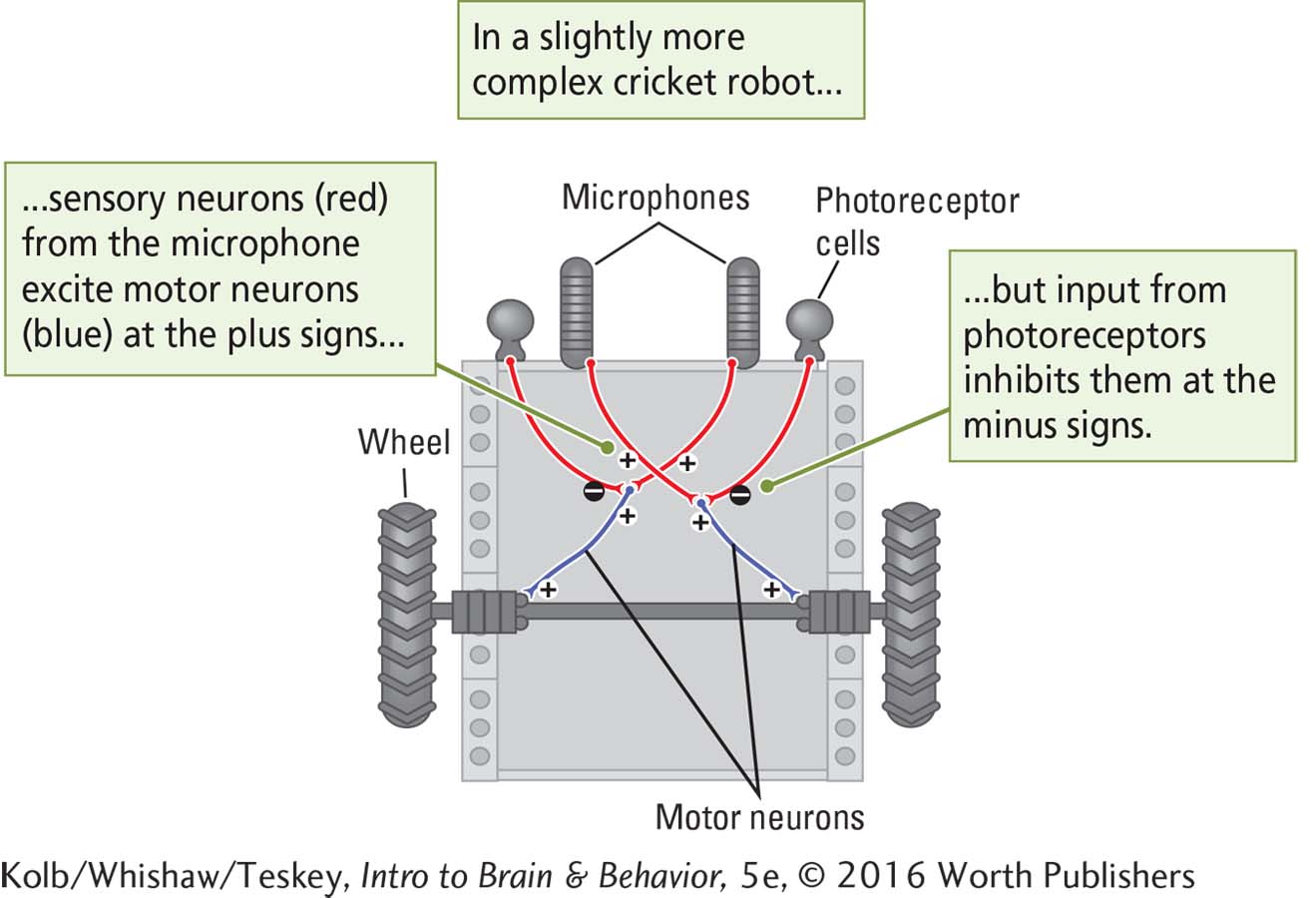
By exciting or inhibiting one another, a neuronal network can detect sensory information and “decide” what kind of motor response to make to that information. To confirm whether they understand how an entire neural network produces behavior, scientists might make a model, such as a robot, intended to function in the same way. Robots, after all, engage in goal-
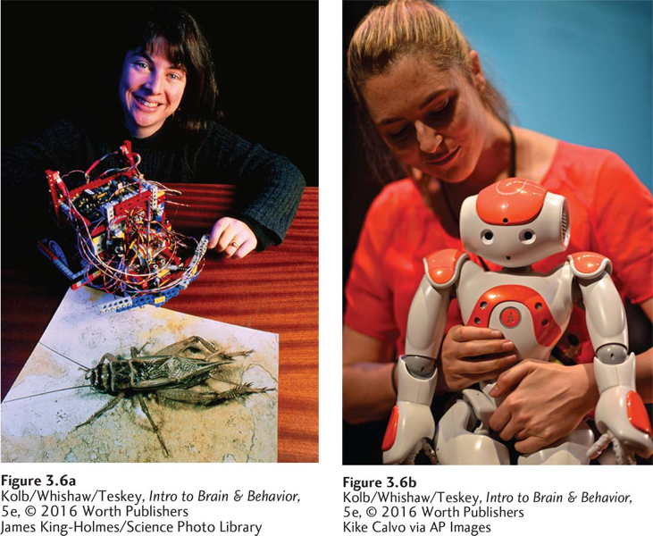
Constructing humanlike machines is one scientific objective of artificial intelligence (AI) research. Barbara Webb’s cricket robot, constructed from Lego blocks, wires, and a motor (Figure 3-6, left), is designed to mimic a female cricket, which listens for the source of a male’s chirping song and travels to it (Reeve et al., 2007). This is the beginning step in constructing intelligent robots (Figure 3-6, right).
In approaching a male, a female cricket must avoid open, well-
The Procedure sections of Experiment 3-1 illustrate some ways that neural inhibition and excitation might produce the cricket robot’s behavior. The Results section confirms that this hypothetical arrangement mimics the functions of sensory and motor neurons and the principle of summating excitatory and inhibitory signals—
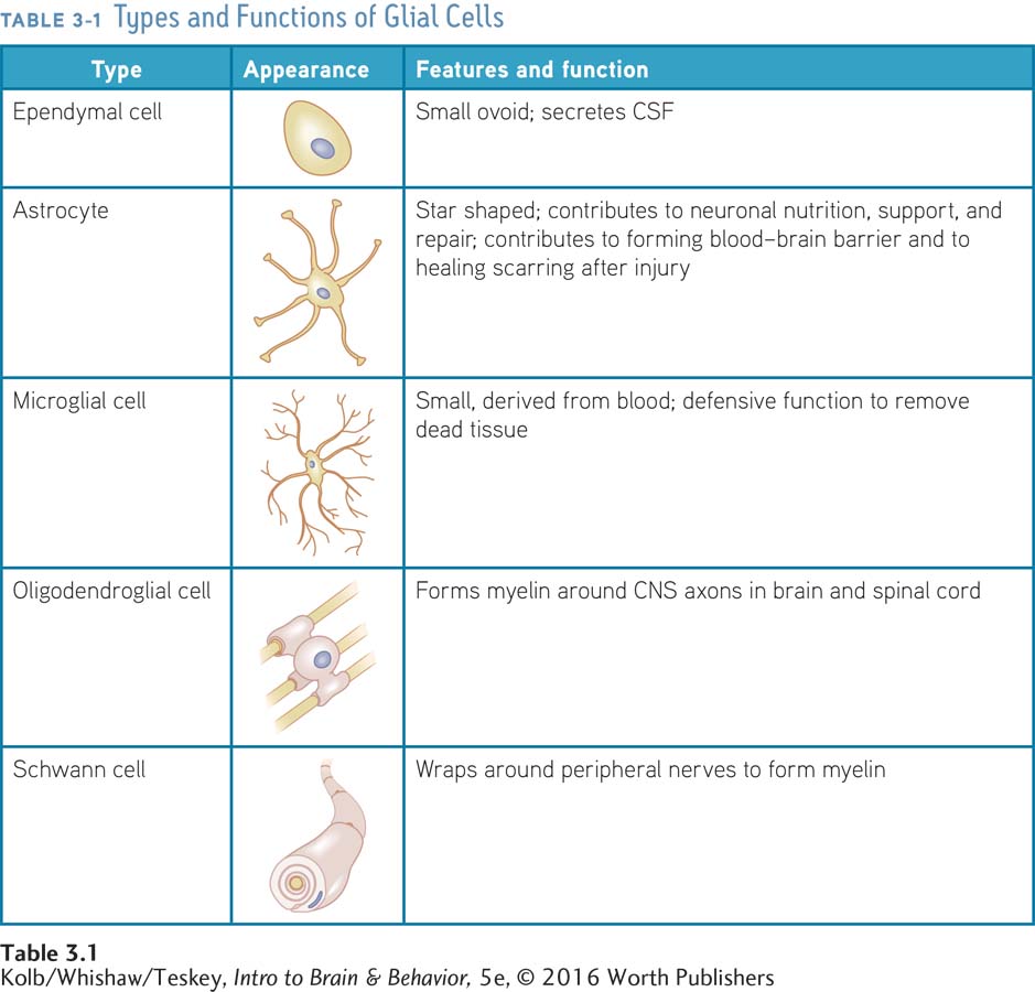
Five Types of Glial Cells
Another group of nervous system cell aids neurons in processing information. Glial cells (from the Greek for glue) are the nervous system’s support cells. The human brain’s estimated 87 billion glial cells reflect the typical nervous system ratio of roughly 1:1 between neurons and glial cells (Herculano-
81
EXPERIMENT 3-1
Question: Can the principles of neural excitation and inhibition control the activity of a simple robot?
Procedure A
If we insert sensory neurons between the microphone for sound detection on each side of this hypothetical robot and the motor on the opposite side, we need only two rules to instruct the female robot to seek out a chirping male cricket.
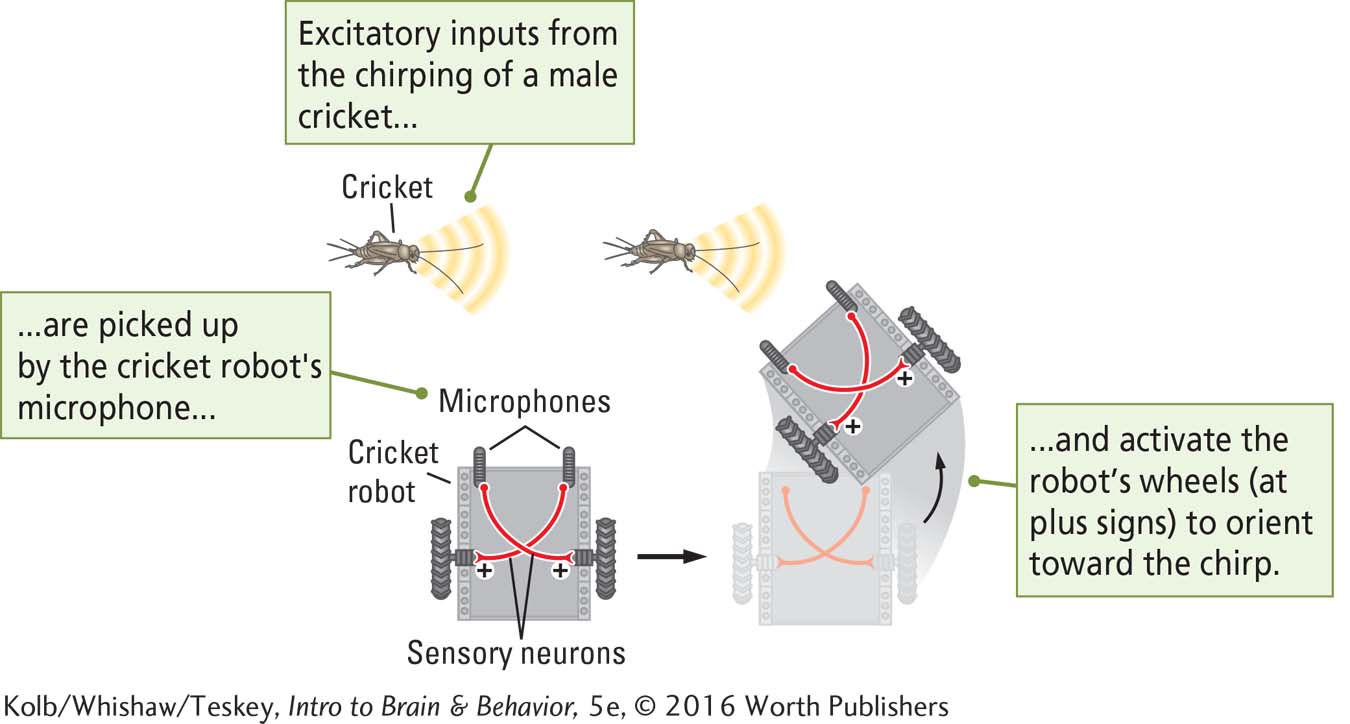
Rule 1 When a microphone detects a male cricket’s song, an excitatory message is sent to the opposite wheel’s motor, activating it so the robot turns toward the cricket.
Rule 2 If the chirp is coming from the robot’s left side, it will be detected as being louder by the microphone on the left, which will make the right wheel turn a little faster, swinging the robot to the left.
Procedure B
We add two more sensory neurons, coming from photoreceptors on the robot. When activated, these light-
Result
This hypothetical arrangement illustrates the biological function of sensory and motor neurons and the neural principle of summating excitatory and inhibitory signals.
Conclusion: A simple robot will operate on the principles of neural excitation and inhibition. More neurons are necessary to make the robot more “intelligent.”
Glia form the fatty coverings around neurons—
Table 3-1 lists the five major types of glia along with their characteristic structures and functions. Glial cells are different from neurons in that new glial cells are produced, and errors in replication can result in abnormal growths: brain tumors. Clinical Focus 3-3, Brain Tumors, describes the results of such uncontrolled glial cell growth.
82
Ependymal Cells
On the walls of the ventricles, the fluid-
Figure 2-9 shows the location of the cerebral aqueduct and the four ventricles.
As CSF flows through the ventricles, it passes through some narrow passages, especially from the cerebral aqueduct into the fourth ventricle, which runs through the brainstem. If these passages are fully or partly blocked, fluid flow is restricted. Because CSF is continuously being produced, this blockage causes a buildup of pressure that begins to expand the ventricles, which in turn push on the surrounding brain.
If such a blockage develops in a newborn infant, before the skull bones are fused, the pressure on the brain is conveyed to the skull, and the baby’s head swells. This condition, called hydrocephalus (literally, water brain), can cause severe intellectual impairment, even death. To treat it, doctors insert one end of a tube, called a shunt, into the blocked ventricle and the other end into a vein, allowing excess CSF to drain into the bloodstream.
83
CLINICAL FOCUS 3-3
Brain Tumors
One day while watching a movie in a neuropsychology class, R. J., a 19-
A few days later, a computed tomography (CT) scan showed a tumor over her left frontal lobe. The tumor was removed surgically, and R. J. returned to classes after an uneventful recovery. Her symptoms have not recurred.
A tumor is an uncontrolled growth of new tissue that is independent of surrounding structures. No region of the body is immune, but the brain is a site for the more than 120 kinds of tumors. They are a common cause of brain cancer in children.
The incidence of brain tumors in the United States is about 20 per 100,000, according to the Central Brain Tumor Registry of the United States (Quinn et al., 2014). In adults brain tumors grow from glia or other supporting cells rather than from neurons, but in infants, tumors may grow from developing neurons. Rate of tumor growth depends on the type of cell affected.
Some tumors are benign, as R. J.’s was, and not likely to recur after removal. Others are malignant, likely to progress, to invade other tissue, and apt to recur after removal. Both kinds can pose a risk to life if they develop in sites from which they are difficult to remove.
The earliest symptoms usually result from increased pressure on surrounding brain structures. They can include headaches, vomiting, mental dullness, changes in sensory and motor abilities, and seizures such as R. J. had. Many symptoms depend on the tumor’s location. The three major types of brain tumors are classified according to how they originate:
Gliomas arise from glial cells. They are slow growing, not often malignant, and relatively easy to treat if they arise from astrocytes. Gliomas that arise from the precursor blast or germinal cells that grow into glia are more often malignant, grow more quickly, and often recur after treatment. U.S. Senator Edward Kennedy was diagnosed with a malignant glioma in 2008 and died a year later. As with R. J., his first symptom was an epileptic seizure.
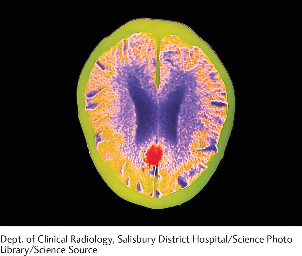
Meningiomas, such as R. J.’s, attach to the meninges and so grow entirely outside the brain, as shown in the accompanying CT scan. These tumors are usually encapsulated (contained), and if the tumor is accessible to surgery, chances of recovery are good.
Metastatic tumors become established when cells from one region of the body transfer to another area (which is what metastasis means). Typically, metastatic tumors are present in multiple locations, making treatment difficult. Symptoms of the underlying condition often first appear when the tumor cells reach the brain.
Treatment for brain tumors is usually surgical, and surgery also remains a main diagnostic tool. Chemotherapy is less successful in treating brain tumors than tumors elsewhere in the body, because the blood–
Recent approaches to treating brain tumors exploit biochemical differences between tumors and healthy tissue, for example to visualize a tumor for surgical removal. Chlorotoxin, a scorpion venom that selectively binds to chloride channels in tumor cell membranes, combined with a fluorescent dye, forms a tumor paint (Butte et al., 2014). Injected into a vein, the paint crosses the blood–
Nonsurgical tumor removal might also replace or aid chemotherapy and radiation therapy. It combines chlorotoxin with a poison to form a drug that kills tumor cells (Wang and Guo, 2015).
Astroglia
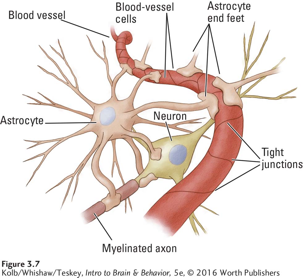
Astrocytes (star-
At the same time, astrocytes contribute to the structure of a protective partition between blood vessels and the brain, the blood–
84
The molecules (smallest units) of these substances are too large to pass between the blood vessel cells unless the blood–
Yet another important function of astrocytes is to enhance brain activity. When you engage in any behavior, whether it’s reading or running, the neuronal network responsible for that behavior requires more fuel in the form of oxygen and glucose. In response to neuron activity, the blood vessels that supply it expand, allowing greater oxygen-
Astrocytes also contribute to healing damaged brain tissue. If the brain is injured by a blow to the head or penetrated by some object, astrocytes form a scar to seal off the damaged area. Although the scar tissue is beneficial in healing the injury, it can also act as a barrier to the regrowth of damaged neurons. One experimental approach to repairing brain tissue seeks to get the axons and dendrites of CNS neurons to grow around or through a glial scar.
Growth factors, described Section 8-2, are chemicals that stimulate and support growth, survival, and perhaps even brain cell plasticity (see Section 14-4).
Microglia
Unlike other glial cells, which originate in the brain, microglia originate in the blood as an offshoot of the immune system and migrate throughout the nervous system, where they make up about 20% of all glial cells. The brain is largely immune privileged, because the blood–
There are several kinds of microglia, which take different shapes depending upon the role they are performing. Microglia engulf any foreign tissue and dead brain cells, an immune process called phagocytosis. When full they take on a distinctive appearance. The stuffed and no-
Because microglia are frontline players in protecting the nervous system and removing its waste, considerable research is directed toward the extent to which microglia are involved in protecting the nervous system from disease. A characteristic of Alzheimer disease, the degenerative brain disorder commonly associated with aging, is the deposit of distinctive bodies called plaques in regions of damage. Microglia may also play a harmful role, consuming inflamed tissue rather than protecting it. They also interact with astrocytes in brain healing. Although small, as their name suggests, microglia play a mighty role in maintaining the brain’s health (Casano & Peri, 2015).
85
More on understanding and treating Alzheimer disease in Section 16-3.
Oligodendroglia and Schwann Cells
Two kinds of glial cells insulate neuronal axons. Like the plastic insulation on electrical wires, myelin prevents adjacent neurons from short-
Oligodendroglia myelinate axons in the brain and spinal cord by sending out large, flat branches that enclose and separate adjacent axons (the prefix oligo- means few and here refers to the fact that these glia have few branches in comparison with astrocytes; see Table 3-1). Schwann cells myelinate axons in the PNS. Each Schwann cell wraps itself repeatedly around a part of an axon, forming a structure somewhat like a bead on a string. In addition to myelination, Schwann cells and oligodendroglia contribute to a neuron’s nutrition and functioning by absorbing chemicals that the neuron releases and releasing chemicals that the neuron absorbs.
Section 4-2, describes how myelin speeds up the neuron’s information flow. Focus 4-2 describes effects of myelin damage.
Glial Cells and Neuron Repair
The multifaceted relations among neurons and glia provide insights into nervous system diseases and into brain injury and recovery from brain injury. Diseases that damage myelin impair a neuron’s ability to send information. The consequences of damage to oligodendroglia and Schwann cells can be as debilitating as damage to neurons themselves. Damage to areas rich in neuron cell bodies can also be different from damage to fiber pathways, because pathway damage includes both neurons and myelin cells.
Glia can also aid in nervous system repair. A deep cut on your body—
Both microglia and Schwann cells participate in repairing damage to the peripheral nervous system. When a PNS axon is cut, it dies back to the cell body, as shown at the top of Figure 3-9. Microglia remove all of the debris left by the dying axon. Meanwhile, the Schwann cells that provided the axon’s myelin shrink and divide to form numerous smaller glial cells along the path the axon formerly took. The cell body then sends out axon sprouts that search for and follow the path formed by the Schwann cells.
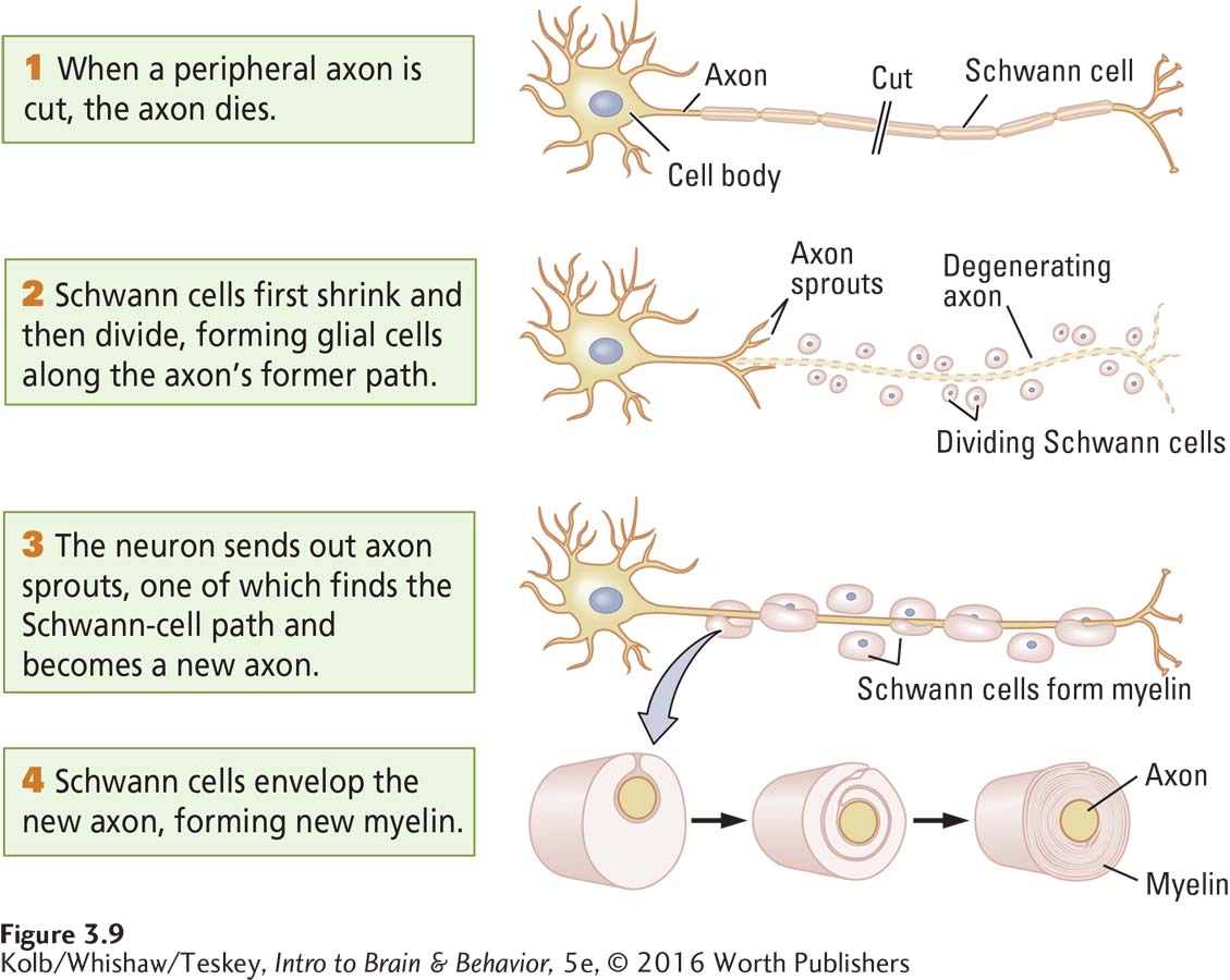
Eventually, one sprout reaches the intended target and becomes the new axon; all other sprouts retract. The Schwann cells envelop the new axon, forming new myelin and restoring function. In the PNS, then, Schwann cells serve as signposts to guide axons to their appropriate end points. Axons can get lost, however, as sometimes happens after surgeons reattach a severed limb. If axons destined to innervate one finger end up innervating another finger instead, the wrong finger will move when a message is sent along that axon.
86
Sections 11-1 and 11-4 detail causes of and treatments for spinal cord injury.
When the CNS is damaged, as happens, for example, when the spinal cord is cut, regrowth and repair do not occur, even though the distance that damaged fibers must bridge is short. The oligodendrocytes that myelinate CNS cells do not behave like PNS Schwann cells to encourage brain repair. They may actually play a role in inhibiting neuron regrowth (Rusielewicz et al., 2014). That recovery should take place in the PNS but not in the CNS is puzzling. Regrowth in the CNS may not occur in part because as neuronal circuits mature, they become exquisitely tuned to mediate individualized behavior and so are protected from the proliferation of new cells or the regrowth of existing cells.
3-1 REVIEW
Cells of the Nervous System
Before you continue, check your understanding.
Question 1
The two classes of nervous system cells are ________, which in humans number around ________, and ________, which in humans number about ________, reflecting the typical ratio.
Question 2
Neurons, the information-
Question 3
The three types of neurons and their characteristic functions are ________, which ________; ________, which ________; and ________, which ________.
Question 4
The five types of glial cells are ________, ________, ________, ________, and ________. Their functions include ________, ________, ________, ________, and ________ neurons.
Question 5
What is the main obstacle to producing a robot with all of the behavioral abilities displayed by a mammal?
Answers appear in the Self Test section of the book.