3-2 Internal Structure of a Cell
What is it about the structure of neurons that generates the remarkable ability to receive, process, store, and send a seemingly limitless amount of information? To answer this question, we must look inside a neuron to see what its components are and understand what they do. Although neurons are minuscule, when we view them with an electron microscope, we find packed inside hundreds of interrelated parts that do the cell’s work.
To a large extent, a cell’s proteins determine its characteristics and functions. Each cell can manufacture thousands of proteins, which variously take part in building the cell and in communicating with other cells. When a neuron malfunctions or contains errors, proteins are implicated and so are involved in many kinds of brain disease. In this section, we explain how the different parts of a cell contribute to protein manufacture, describe what a protein is, and detail some functions of proteins.
Water, salts, and ions play prominent parts in the cell’s functions, as you will learn in this and the next few chapters. If you already understand the structure of water and you know what a salt is and what ions are, read on. If you prefer a brief chemistry review first, turn to The Basics: Chemistry Review, on pages 88–89.
The Cell as a Factory
87
We began Section 3-1 by comparing a cell to a miniature factory, with work centers that cooperate to make and ship the cell’s products—
Figure 3-10 displays many external and internal cellular components. A factory’s outer wall separates it from the rest of the world and affords some security. Likewise, a cell’s double-
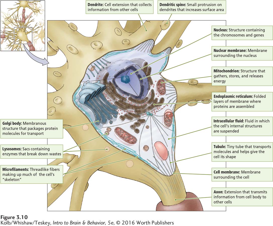
Very few substances can enter or leave a cell spontaneously, because the cell membrane is virtually impermeable (impenetrable). Some proteins made by the cell are embedded in the cell membrane, where they facilitate the transport of substances into and out of the cell. These proteins thus serve as the cellular factory’s gates.
88
THE BASICS
Chemistry Review
The smallest unit of a protein or any other chemical substance is the molecule. Molecules and the even smaller atoms of elements that constitute them are the cellular factory’s raw materials.
Elements, Atoms, and Ions
Chemists represent each element, a substance that cannot be broken down into another substance, by a symbol—
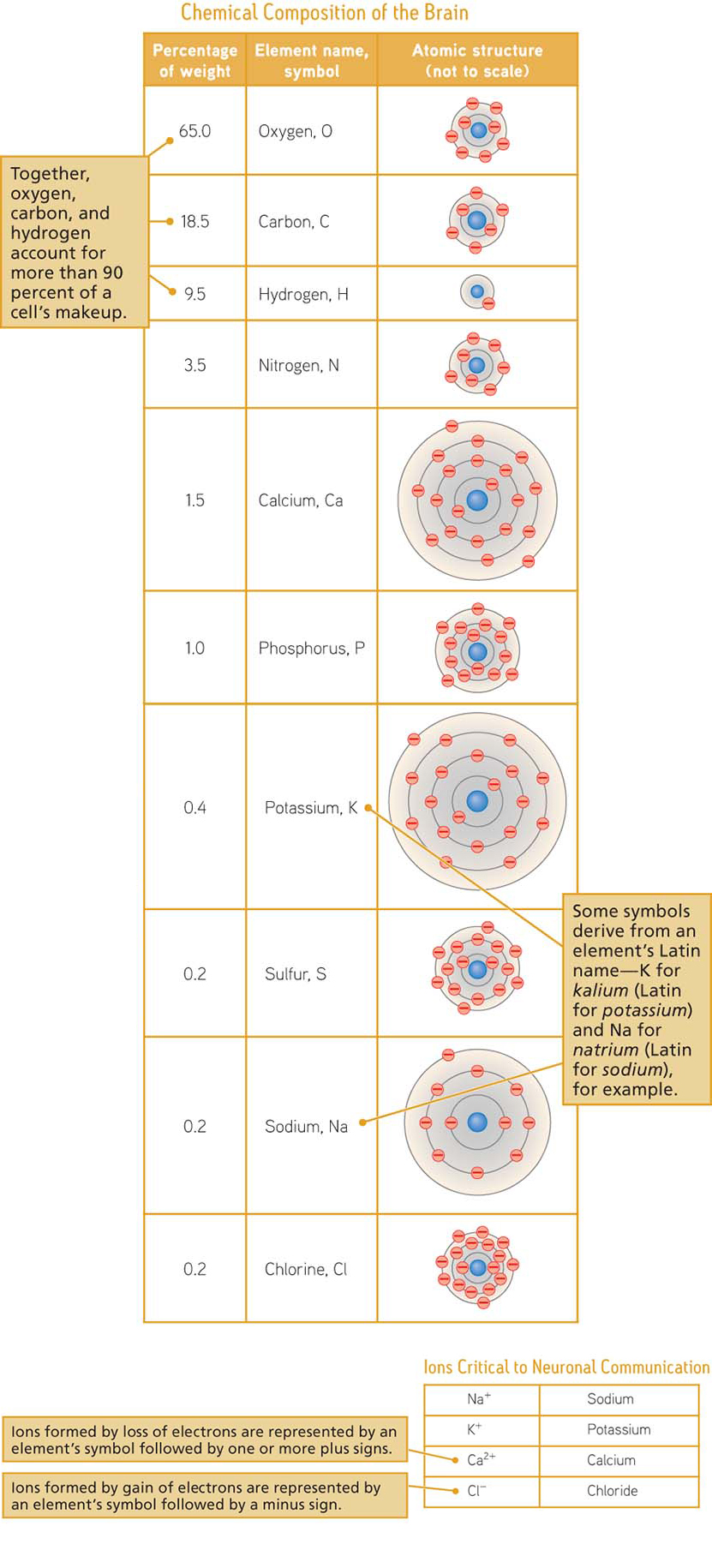
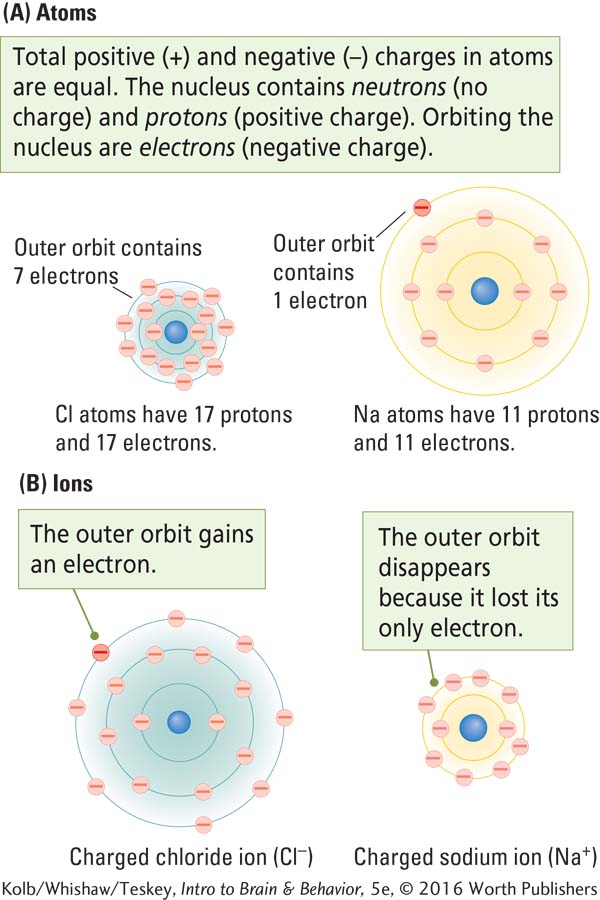
The smallest quantity of an element that retains the properties of that element is an atom. Ordinarily, as shown opposite in part A of the figure Ion Formation, atoms are electrically neutral: their total positive and negative charges are equal.
Atoms of chemically reactive elements such as sodium and chlorine can easily lose or gain negatively charged particles, or electrons. When an atom gives up electrons, it becomes positively charged; when it takes on extra electrons, it becomes negatively charged, as illustrated in part B of Ion Formation. Either way, the charged atom is now an ion. Ions’ positive or negative charges allow them to interact. This property is central to cell function.
89
Molecules: Salts and Water
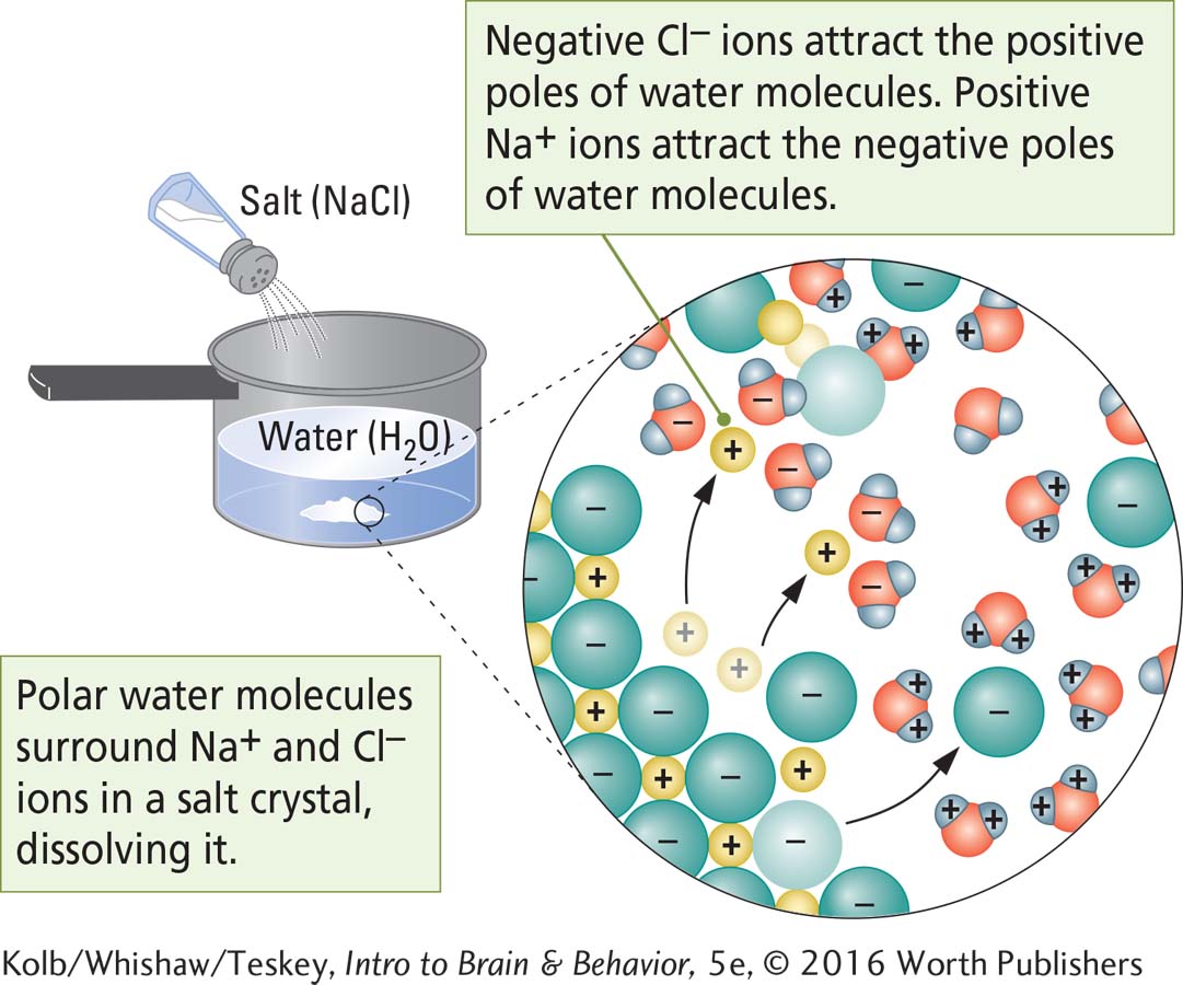
Salt crystals form bonds via the electrical attraction between ions. The formula for table salt, NaCl (sodium chloride), means that this molecule consists of one sodium ion and one chloride ion. KCl, the formula for the salt potassium chloride, is composed of one potassium ion (K+) and one chloride ion (Cl–).
Atoms bind together to form molecules, the smallest units of a substance that contain all of its properties. A water molecule (H2O) is the smallest unit of water that retains the properties of water. Breaking down water any further would release its component elements, the gases hydrogen and oxygen. The symbol H2O indicates that a water molecule is the union of two hydrogen atoms and one oxygen atom.
Ionic bonds hold salt molecules together, but the atoms of water molecules share electrons and electron sharing is not equal: H electrons spend more time orbiting the O atom than orbiting each H atom. As shown above in part A of Chemistry of Water, this structure gives the oxygen region of the water molecule a slight negative charge and leaves the hydrogen regions with a slight positive charge. Like atoms, most molecules are electrically neutral, but water is polar: it carries opposite charges on opposite ends, just as Earth does at the North and South Poles.
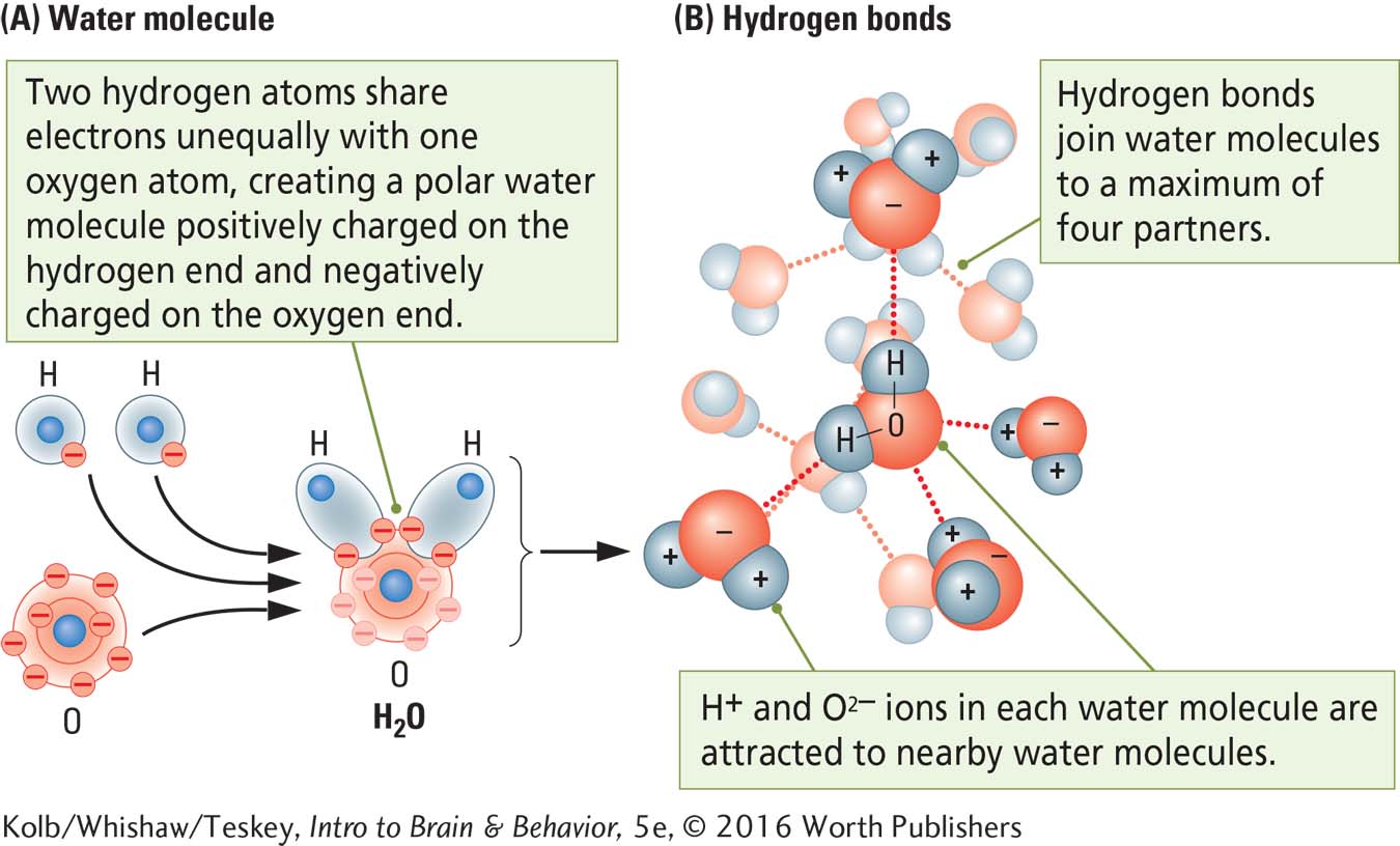
Because water molecules are polar, they are attracted to other electrically charged substances and to one another. Part B of Chemistry of Water illustrates this attracting force, called a hydrogen bond. Hydrogen bonding enables water to dissolve electrically neutral salt crystals into their component ions. Salts thus cannot retain their shape in water: they dissolve. As illustrated in the figure Salty Water, the polar water molecules muscle their way into the Na+ and Cl– lattice, surrounding and separating the ions.
Essentially, it is salty water that bathes our brain cells, provides the medium for their activities, supports their communications, and constitutes the brain’s CSF. Sodium chloride and many other dissolved salts, including KCl (potassium chloride) and CaCl2 (calcium chloride) are among the constituents of the brain’s salty water.
90
Although neurons and glia appear to be packed tightly together, like all cells, they are separated by extracellular fluid composed mainly of water with dissolved salts and many other chemicals. A similar intracellular fluid is found inside a cell. What’s important is the cell membrane’s relative impermeability, which ensures that concentrations of substances inside and outside the cell are different.
In the CNS, the extracellular fluid is CSF.
Within the cell shown in Figure 3-10 are membranes that surround its organelles, similar to the work areas demarcated by a factory’s interior walls. Each organelle membrane is also relatively impermeable and so concentrates needed chemicals while keeping out unneeded ones.
The prominent nuclear membrane surrounds the cell’s nucleus. Within the nucleus the genetic blueprints for the cell’s proteins are stored, copied, then sent to the “factory floor,” the endoplasmic reticulum. The ER is an extension of the nuclear membrane, and here the cell’s protein products are assembled in accordance with instructions from the nucleus. Once those proteins are assembled, many are packaged and sent throughout the cell. The Golgi bodies are “mailrooms,” where proteins are wrapped, addressed, and shipped.
Other cell components are tubules of several kinds. Some (microfilaments) reinforce the cell’s structure; others aid in the cell’s movements. Still others (microtubules) form the transportation network that carries proteins to their destinations, much as roads allow a factory’s trucks and forklifts to deliver goods to their destinations.
Two other important parts of the cellular factory shown in Figure 3-10 are the mitochondria (sing. mitochondrion), the cell’s power plants, which supply its energy needs, and lysosomes, vesicles that transport incoming supplies and remove and store wastes. Interestingly, more lysosomes are found in old cells than in young ones. Cells apparently have trouble disposing of all of their garbage, just as societies do.
Cell Membrane: Barrier and Gatekeeper
The cell membrane separates the intracellular from the extracellular fluid, allowing the cell to function as an independent unit. The membrane’s double-
91
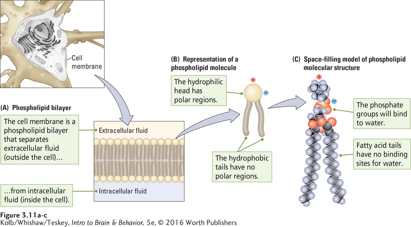
The cell membrane also regulates the differing concentrations of salts and other chemicals on its inner and outer sides. This regulation is important because, if its concentrations of chemicals are unbalanced, the cell will not function normally. What properties of a cell membrane allow it to regulate water and salt concentrations? One property is its special molecular construction. These molecules, called phospholipids, are named for their structure, shown close up in Figure 3-11B.
Figure 3-11C shows a space-
The polar head and the nonpolar tails are the underlying reasons that a phospholipid molecule can form cell membranes. The heads are hydrophilic (Greek hydro, meaning water, and philia, meaning love) and so are attracted to one another and to polar water molecules. The nonpolar lipid tails have no such attraction for water. They are hydrophobic, or water hating (from the Greek word phobia, meaning fear).
Quite literally, then, the head of a phospholipid loves water and the tails hate it. To avoid water, the tails of phospholipid molecules point toward each other, and the hydrophilic heads align with one another and point outward to the watery intracellular and extracellular fluid. In this way, the cell membrane consists of a bilayer (two layers) of phospholipid molecules (see Figure 3-11A).
The bilayer cell membrane is flexible even as it forms a formidable barrier to a wide variety of substances. It is impenetrable to intracellular and extracellular water, because polar water molecules cannot pass through the hydrophobic tails on the membrane’s interior. Ions in the extracellular and intracellular fluid also cannot penetrate this membrane, because they carry charges and thus cannot pass by the polar phospholipid heads. In fact, only a few small molecules, such as oxygen (O2), carbon dioxide (CO2), and the sugar glucose, can traverse a phospholipid bilayer.
The Nucleus and Protein Synthesis
In our factory analogy, the nucleus is the cell’s executive office, where the blueprints for making proteins are stored, copied, and sent to the factory floor. These blueprints are called genes, segments of DNA that encode the synthesis of particular proteins. Genes are contained within the chromosomes, the double-
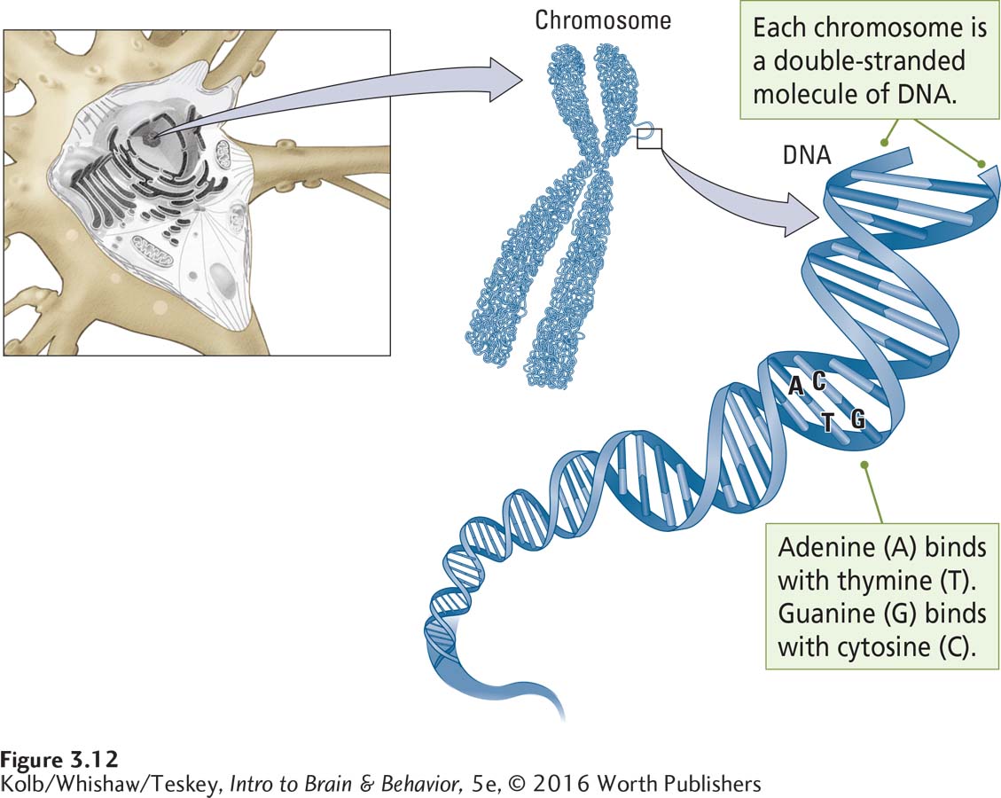
The chromosomes have been likened to a book of blueprints. Each chromosome contains thousands of genes. Each gene is the blueprint, or code, for making one protein. The location of the chromosomes in the cell nucleus, the appearance of a chromosome, and the structure of the DNA within a chromosome are shown in Figure 3-12.
Chromosome means colored body; they are so named because chromosomes can be readily stained with certain dyes.
This static picture of chromosomes does not represent the way they look in living cells. Videos of the cell nucleus show that chromosomes are constantly changing shape and moving in relation to one another, jockeying to occupy the best locations within the nucleus. By changing shape, chromosomes expose different genes to the surrounding fluid, thus allowing the gene to begin the process of making a protein.
92
A human somatic (body) cell has 23 pairs of chromosomes, or 46 chromosomes in all (in contrast, the 23 chromosomes within a reproductive cell are not paired). Each chromosome is a double-
Each strand possesses a variable sequence of four nucleotide bases, the constituent molecules of the genetic code: adenine (A), thymine (T), guanine (G), and cytosine (C). Adenine on one strand always pairs with thymine on the other, whereas guanine on one strand always pairs with cytosine on the other. The two strands of the DNA helix are bound together by the attraction between the two bases in each pair, as illustrated in Figure 3-12. Sequences of hundreds of nucleotide bases within the chromosomes spell out the genetic code. Scientists represent this code by the letters of the nucleotide bases, for example ATGCCG and so forth.
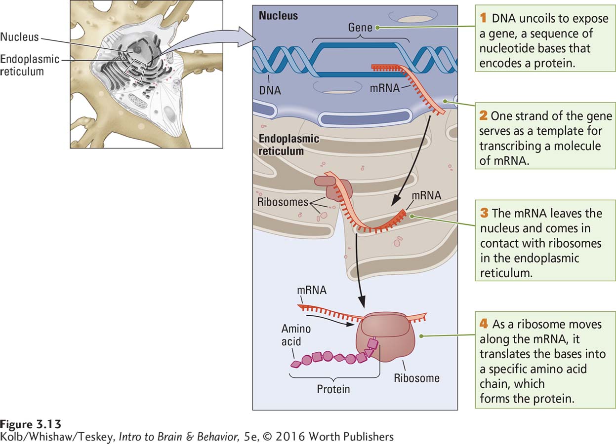
A gene is a segment of a DNA strand. A gene’s code is its sequence of thousands of nucleotide bases. Much as a sequence of letters spells out a word, the sequence of ACTG base pairs spells out the order in which amino acids, the constituent molecules of proteins, should be assembled to construct a certain protein. To begin to make a protein, the appropriate gene segment of the DNA strands first unwinds to expose its bases. The exposed sequence of nucleotide bases on one of the DNA strands then serves as a template to attract free-
93
The Endoplasmic Reticulum and Protein Manufacture
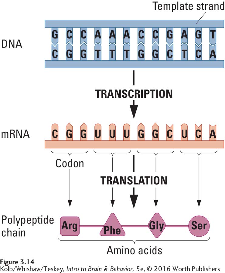
RNA produced through transcription is much like a single strand of DNA except that the base uracil (U), which also is attracted to adenine, takes the place of thymine. The transcribed strand of RNA is called messenger RNA (mRNA) because it carries the protein code (the message) out of the nucleus to the endoplasmic reticulum, where proteins are manufactured.
Steps 3 and 4 in Figure 3-13 show that the ER consists of membranous sheets folded to form numerous channels. A distinguishing feature of the ER is that it may be studded with ribosomes, protein structures that act as catalysts to facilitate the building of proteins. When an mRNA molecule reaches the ER, it passes through a ribosome, where its genetic code is read. In this process of translation, a particular sequence of nucleotide bases in the mRNA is transformed into a particular sequence of amino acids. Transfer RNA (tRNA) assists in translating nucleotide bases into amino acids.
As shown in Figure 3-14, each group of three consecutive nucleotide bases along an mRNA molecule encodes one particular amino acid. These sequences of three bases are called codons. For example, the codon uracil, guanine, guanine (UGG) encodes the amino acid tryptophan (Trp), whereas the codon uracil, uracil, uracil (UUU) encodes the amino acid phenylalanine (Phe). The sequence of codons on the mRNA strand determines the sequence of the resulting amino acid chain.
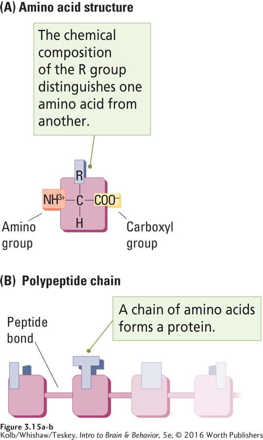
Humans utilize 20 different amino acids, all structurally similar, as illustrated in Figure 3-15A. Each consists of a central carbon atom (C) bound to a hydrogen atom (H), an amino group (NH3+), a carboxyl group (COO–), and a side chain (represented by the letter R). The side chain varies in chemical composition from one amino acid to another. Each amino group (NH3+) is bound to the carboxyl group (COO–) of the adjacent amino acid by a peptide bond, which gives amino acid chains their alternative name, polypeptide chain (Figure 3-15B).
Just as a remarkable number of words can be made from the 26 letters of the English alphabet, a remarkable number of polypeptide (meaning many peptides) chains can be made from the 20 amino acids. These amino acids can form 400 (20 × 20) dipeptides (two-
In summary, the information flow driven by the genetic code is conceptually quite simple: a gene (portion of a DNA strand) is transcribed into a strand of mRNA, and ribosomes translate the mRNA into a molecular chain of amino acids, a polypeptide chain. Thus the sequence of events in building a protein:
DNA → mRNA → protein
Proteins: The Cell’s Product
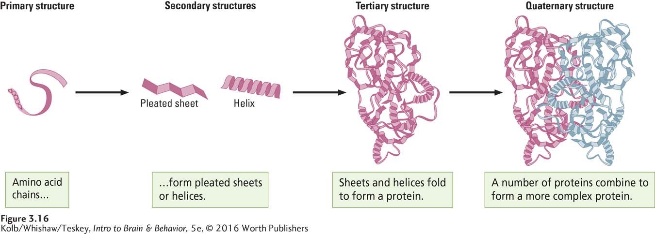
A polypeptide chain and a protein are related, but they are not the same. The relation is analogous to the relation between a length of ribbon and a bow that can be made from the ribbon. Long polypeptide chains have a strong tendency to twist into a helix (a spiral) or to form pleated sheets, which in turn have a strong tendency to fold together to form more complex shapes, as shown in Figure 3-16. A protein is a folded-
Any one neuron contains as many as 20,000 genes, and it can, in principle, produce as many as 20,000 different protein molecules. The number of proteins that can ultimately be produced by a neuron is far larger than the number of its genes, however. Although each gene codes for one protein, a protein can be cleaved into pieces—
94
A protein’s shape and its ability to change shape and to combine with other proteins are central to its function. Through their shapes and changes in shape, proteins can combine with other proteins in chemical reactions. They can modify the length and shape of other proteins and so act as enzymes. Proteins embedded in a cell membrane form passageways called channels, gates, and pumps that regulate the flow of substances through the membrane. And proteins can be exported through the axon terminal to travel to other cells and so act as messenger molecules.
Golgi Bodies and Microtubules: Protein Packaging and Shipment
Getting proteins to the right destination is the task of cellular components that package, label, and ship them. These components operate much like a postal or shipping service.
To reach their appropriate destinations, protein molecules that have been synthesized in the cell are wrapped in membranes and marked with addresses to indicate where they are to go. This wrapping and labeling take place in the organelles called Golgi bodies. The packaged proteins are then loaded onto motor molecules that move along the many microtubules radiating through the cell, carrying each protein to its destination. Protein export is illustrated in Figure 3-17.
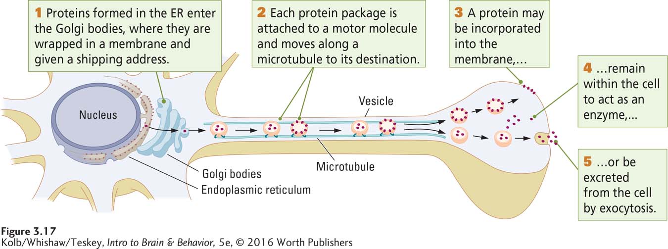
If a protein is destined to remain within the cell, it is unloaded into the intracellular fluid. If it is to be incorporated into the cell membrane, it is carried to the membrane, where it inserts itself. Some proteins are expelled from the cell. In this process, called exocytosis, the membrane, or vesicle, in which the protein is wrapped fuses with the cell membrane, and the protein is excreted into the extracellular fluid. The roles proteins play when embedded in the cell membrane or exported from the cell are central to understanding how neurons process information and determine behavior.
Crossing the Cell Membrane: Channels, Gates, and Pumps
95
Proteins embedded in the cell membrane serve many functions. One is transporting substances across the membrane. We now consider how three such membrane proteins—
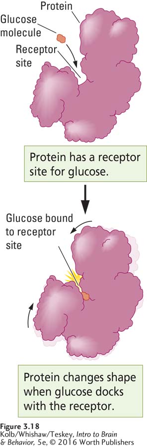
A protein’s shape and its ability to change shape both derive from the precise amino acid sequence that composes the protein molecule. Some proteins change shape when other chemicals bind to them; others change shape as a function of temperature; and still others change shape in response to changes in electrical charge. The protein molecule’s ability to change shape is analogous to a lock in a door. When a key of the appropriate size and shape is inserted into the lock and turned, the locking device activates and changes shape, allowing the door to be closed or opened.
Such a shape-
Some membrane proteins form channels through which substances can pass. Different-
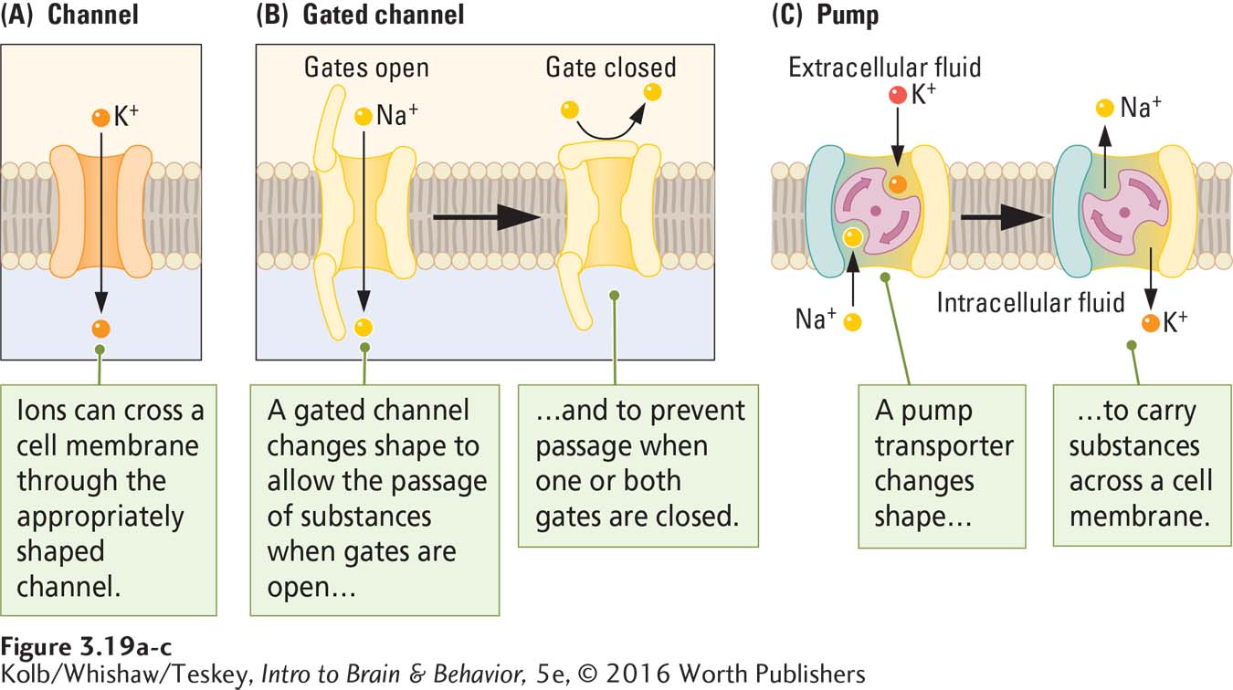
Figure 3-19B shows a protein molecule that acts as a gate to regulate the passage of substances. Like the protein in Figure 3-18, gates change their shape in response to some trigger. The protein allows substances to pass through when its shape forms a channel and prevents passage when its shape leaves the channel closed.
Changes in protein shape can also allow it to act as a pump. Figure 3-19C shows a protein that changes its shape to pump Na+ and K+ across the membrane, exchanging the Na+ on one side for the K+ on the other.
Channels, gates, and pumps play an important role in allowing substances to enter and leave a cell. This passage of substances is critical in explaining how neurons send messages. Chapter 4 explores how neurons use electrical activity to communicate.
96
3-2 REVIEW
Internal Structure of a Cell
Before you continue, check your understanding.
Question 1
The constituent parts of the cell include the __________,__________,__________,__________,__________, and __________.
Question 2
The product of the cell is __________. They serve many functions, including acting at the cell membrane as__________, __________, and __________ to regulate movement of substances across the membrane.
Question 3
The basic sequence of events in building a protein is that __________ makes __________ makes __________.
Question 4
Once proteins are formed in the __________, they are wrapped in membranes by __________ and transported by __________ to their designated sites in the neuron or its membrane or exported from the cell by __________.
Question 5
Why is a cell more than a protein factory?
Answers appear in the Self Test section of the book.