4-4 Into the Nervous System and Back Out
The nervous system allows us to respond to afferent sensory stimuli by detecting them and sending messages about them to the brain. The brain interprets the information, triggering efferent responses that contract muscles and produce behavior. Until now, we have been dealing only with the middle of this process—
To fill in the missing pieces, we explain how a sensory stimulus initiates a nerve impulse and how a nerve impulse produces a muscular contraction. Once again, ion channels are vitally important, but in muscles, the channels are different from those described so far.
How Sensory Stimuli Produce Action Potentials
We receive information about the world through bodily sensations (touch and balance), auditory sensations (hearing), visual sensations (sight), and chemical sensations (taste and olfaction). Each sensory modality has one or more separate functions. In addition to touch, for example, the body senses include pressure, joint sense, pain, temperature, and itch. Receptors for audition and balance are modified touch receptors. The visual system has receptors for light and for colors. And taste and olfactory senses respond to a plethora of chemical compounds.
For detail on how sensory receptors transduce external energy into action potentials:
Hearing, Section 10-1
Sensation and perception, Section 9-1
Smell and taste, Section 12-2
Touch, pain, and balance, Section 11-4
Vision, Section 9-2
To process all these varied sensory inputs requires a remarkable array of sensory receptors. But in all our sensory systems, the neurons related to these diverse receptors have one thing in common: conduction of information begins at ion channels. They initiate the chain of events that produces a nerve impulse.
An example is touch. Each hair on the human body allows us to detect even a very slight displacement. You can demonstrate this sensitivity to yourself by selecting a single hair on your arm and bending it. If you are patient and precise in your experimentation, you will discover that some hairs are sensitive to displacement in one direction only, whereas others respond to displacement in any direction. What enables this finely tuned sensitivity?
133
The base of each hair is wrapped in a dendrite of a touch neuron. When you bend a hair or otherwise mechanically displace it, the encircling dendrite is stretched (Figure 4-25). The displacement opens stretch-
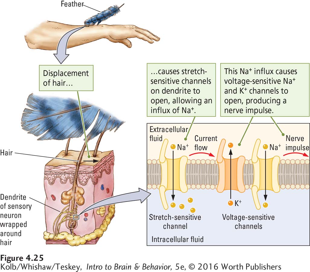
Other kinds of sensory receptors have similar mechanisms for transducing (transforming) the energy of a sensory stimulus into nervous system activity. When displaced, the hair receptors that provide information about hearing and balance likewise activate stretch-
How Nerve Impulses Produce Movement
What happens at the end of the neural journey? After sensory information has traveled to the brain and been interpreted, how does the brain generate output—
Motor neurons send nerve impulses to synapses on muscle cells. These synapses are instrumental in making the muscle contract. Each motor neuron axon makes one or a few synapses with its target muscle, synapses similar to those neurons make with one another Figure 4-26A. The axon terminal contacts a specialized area of the muscle membrane called an end plate. Onto the muscle’s end plate the axon terminal releases the chemical transmitter acetylcholine.

Acetylcholine does not enter the muscle but rather attaches to transmitter-
Sections 5-2 and 5-3 describe the varieties of chemical transmitters and how they function.
The transmitter-
134
CLINICAL FOCUS 4-4
ALS: Lou Gehrig’s Disease
In 1869, French physician Jean-
Gerhig played on many World Series championship teams, but ALS sapped his strength, forcing him to retire from baseball at age 36. Amyotrophic means muscle weakness; lateral sclerosis means hardening of the lateral spinal cord. Gehrig’s condition deteriorated rapidly. He died 2 years later. ALS strikes most commonly at age 50 to 75, although its onset can be as early as the teenage years.
While death often occurs within 5 years of diagnosis, internationally renowned theoretical physicist and cosmologist Stephen Hawking is a notable exception. Diagnosed at age 21, Hawking has a rare early-
Roughly 10 percent of people with ALS have a family history of the disorder. About 5000 new cases are reported in the United States each year. ALS is due primarily to the death of spinal motor neurons but can affect brain neurons as well in some cases.
ALS typically begins with general weakness, at first in the throat or upper chest and in the arms and legs. Gradually, walking becomes difficult and falling common. ALS does not usually affect any sensory systems, cognitive functions, bowel or bladder control, or even sexual function. Even as his motor neurons continue to die, Stephen Hawking’s mind-
At present, no cure for ALS exists, although some newly developed drugs appear to slow its progression and offer some hope for future treatments. In 2014 the ALS Ice Bucket Challenge appeared on YouTube to promote awareness of ALS and encourage donations to research. The Challenge went viral.

Should the acetylcholine receptors on muscle end plates be blocked, acetylcholine released from the motor neuron cannot properly exert its depolarizing effect. This prevents muscular contraction in conditions such as the autoimmune disease myasthenia gravis. In affected individuals the thymus, an immune system gland that normally produces antibodies to foreign material like viruses, makes antibodies to the acetylcholine receptors on muscles, causing weakness and fatigue (Figure 4-27).
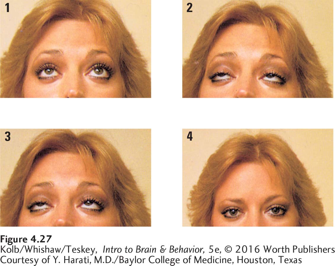
Unlike MS, another autoimmune disease, myasthenia gravis is usually well controlled by treatments including drugs that suppress the immune system or inhibit acetylcholine breakdown, extending the time it can act, and by removal of the thymus gland (thymectomy).
The actions of membrane channels can explain a wide range of neural events. Some channels generate the transmembrane charge. Others mediate graded potentials. Still others trigger the action potential. Sensory stimuli activate channels on neurons to initiate a nerve impulse, and the nerve impulse eventually activates channels on motor neurons to produce muscle contractions.
These various channels and their different functions evolved over a long time in the same way that new species of animals and their behaviors evolve. We have not described all the different ion channels that neural membranes possess, but you will learn about some additional ones in subsequent chapters.
4-4 REVIEW
135
Into the Nervous System and Back Out
Before you continue, check your understanding.
Question 1
Different sensory stimuli initiate nerve impulses for each __________ in a similar way.
Question 2
A(n) __________ membrane contains a mechanism for transducing sensory energy into changes in ion channels. In turn, the channels allow ion flow to alter the membrane voltage to the point __________ that channels open, initiating a nerve impulse.
Question 3
Sensory stimuli activate ion channels to initiate a nerve impulse that activates channels on __________ neurons, which in turn contract __________.
Question 4
In myasthenia gravis, a(n) __________ disease, the thymus gland produces antibodies to __________ receptors on muscles, causing weakness and fatigue.
Question 5
Why have so many kinds of ion channels evolved on cell membranes?
Answers appear in the Self Test section of the book.