SUMMARY
318
9-
Sensory systems allow animals, including ourselves, to adapt. Animals adapted to different environments vary widely in their sensory abilities. What is distinctive about humans is the extent to which we can transform sensations into perceptual information to mediate aspects of language, music, and culture. For each sense, mammals represent the world in topographic maps that form neural–
9-
Like all sensory systems, vision begins with receptor neurons. The visual photoreceptors (rods and cones) at the back of the eye in the retina transduce the physical energy of light waves into neural activity.
Rods are sensitive to dim light. Cones, which are sensitive to bright light, mediate color vision. Each of the three cone types is maximally sensitive to a different wavelength—
Retinal ganglion cells receive input from photoreceptors through bipolar cells and send their axons out from the retinas to form the optic nerve. P ganglion cells receive input mostly from cones and convey information about color and fine detail. M cells receive input from rods and convey information about luminance and movement but not color.
The optic nerve forms two distinct major routes into the brain. The geniculostriate pathway synapses first in the thalamic LGN nucleus, then in V1. The tectopulvinar pathway synapses first in the midbrain’s tectum (superior colliculus), then in the pulvinar of the thalamus, and finally in the temporal and parietal visual cortex areas. A few optic nerve fibers also form the retinohypothalamic tract, which functions in part to control circadian rhythms.
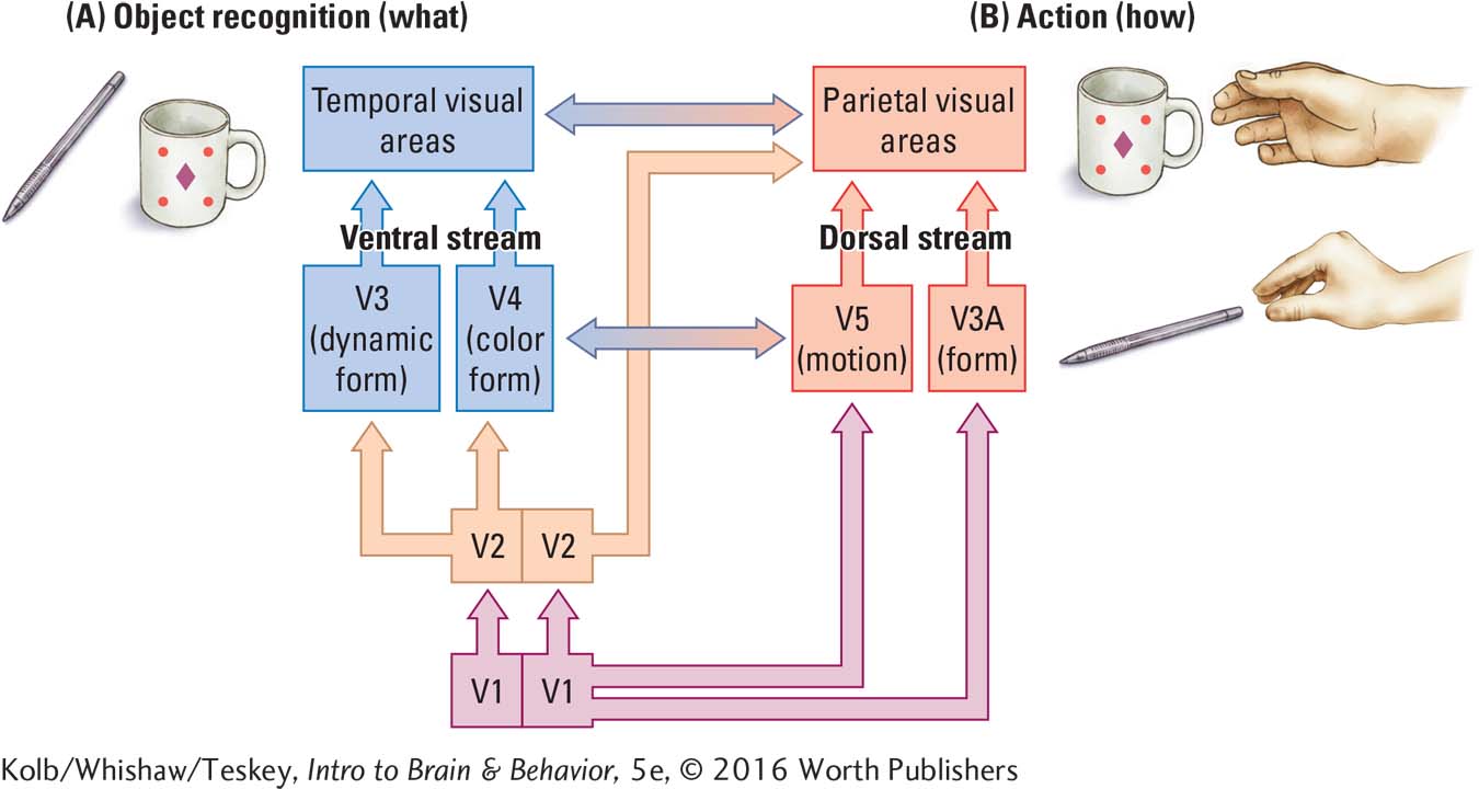
Among the visual regions in the occipital cortex, V1 and V2 carry out multiple functions; the remaining regions (V3, V3A, V4, and V5) are specialized. Visual information flows from the thalamus to V1 and V2, then divides to form the visual stream pathways. The unconscious dorsal stream aids in guiding movements visually, whereas the conscious ventral stream aids in visual object perception.
9-
At each step along the visual pathways, neuronal activities are distinctly different; it is the summed neural activity in all regions that produces our visual experience. Each functional column in the cortical visual regions is about 0.5 mm in diameter and extends to the depth of the cortex. The visual system cortical columns are specialized for processes such as analyzing line orientation or comparing similar shapes as complex as faces.
9-
Neurons in the ventral stream are selective for aspects of shape. Those in the visual cortex are maximally responsive to lines of different orientations. Upstream, cells in the inferior temporal cortex are responsive to shapes, some abstract, and in other cases, to concrete forms as complex as hands or faces.
Cones in the retina are maximally responsive to different light wavelengths, roughly corresponding to colors we perceive as green, blue, and red. At the next level, RGCs’ center–
Color-
9-
Upon entering the brain, information from the left and right visual fields proceeds on the optic nerve to the brain’s right and to its left sides, respectively. As a result of these contralateral connections, damage to the visual areas on one side of the brain results in visual disturbance in both eyes, because half of each retina’s visual field is represented on each side of the brain.
Specific visual functions are localized to different brain regions, so local damage results in the loss of a particular function. Damage to region V4 produces a loss of color constancy, for example; damage to regions in the parietal cortex inhibits the contralateral hand’s grasping ability.
As summarized in the illustration, the visual streams perform distinct functions: (A) object recognition (the what) in the ventral stream and (B) visual action (the how) in the dorsal stream. We are largely unconscious of the dorsal stream’s ongoing online analyses, that allow us to make accurate movements in relation to objects.