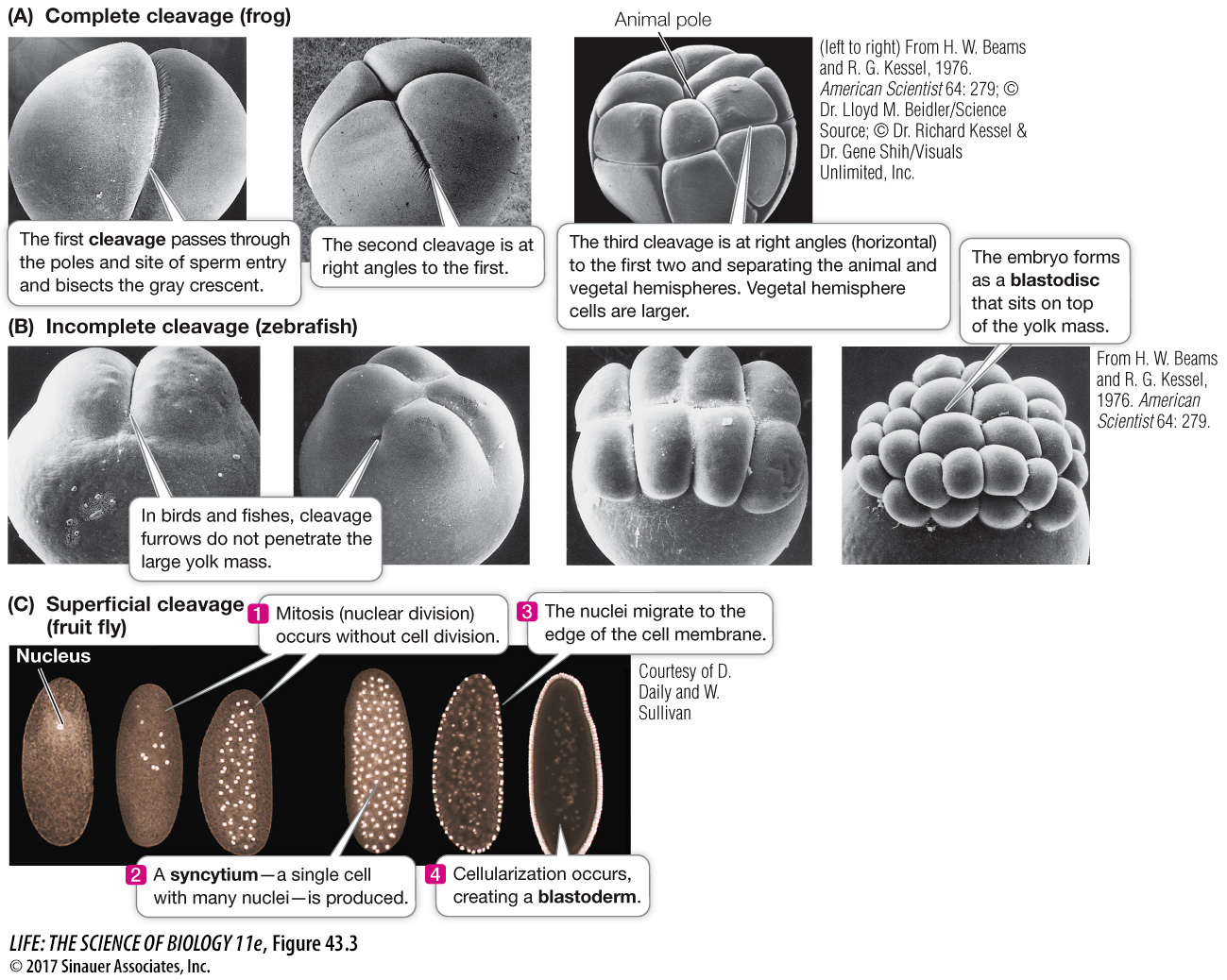
Figure 43.3 Some Patterns of Cleavage Differences in patterns of early embryonic development reflect differences in the way the egg cytoplasm is organized. (A) The frog is a model organism representing complete cleavage in these scanning electron micrographs (SEMs). (B) SEMs of zebrafish embryos illustrate incomplete cleavage, in which the large yolk mass limits the planes of cleavage. (C) Nuclear staining reveals the syncytial nuclei characteristic of the early embryo of a fruit fly. These nuclei migrate to the periphery. Cleavage furrows then move inward to separate the nuclei into individual cells, forming the blastoderm.