image
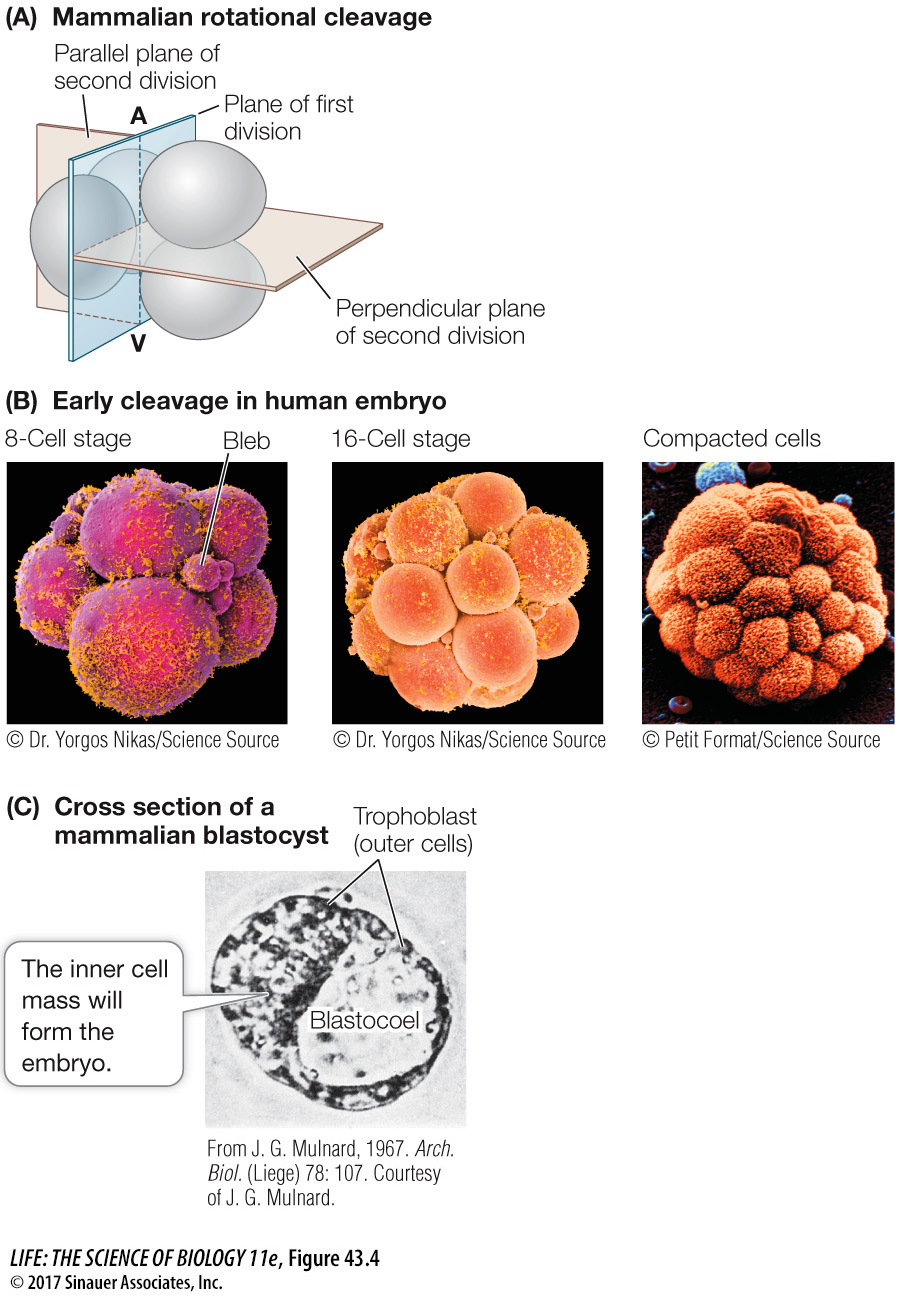
Figure 43.4 Becoming a Blastocyst (A) Mammals have rotational, complete cleavage, in which the plane of the first cleavage is parallel to the animal–vegetal (A–V) axis, but the second cell division involves two planes (beige) at right angles to each other. (B) Scanning electron micrographs (color added) of early cleavage (leading to the formation of the blastocyst) in a human embryo. The cells’ outer surfaces are covered with cilia (bright yellow). The small spheres, or “blebs,” of cytoplasmic material, prominent at the 8-cell stage, disintegrate as cleavage progresses. (C) Seen in cross section under a light microscope, a mammalian blastocyst consists of an inner cell mass adjacent to a fluid-filled blastocoel and surrounded by trophoblast cells.