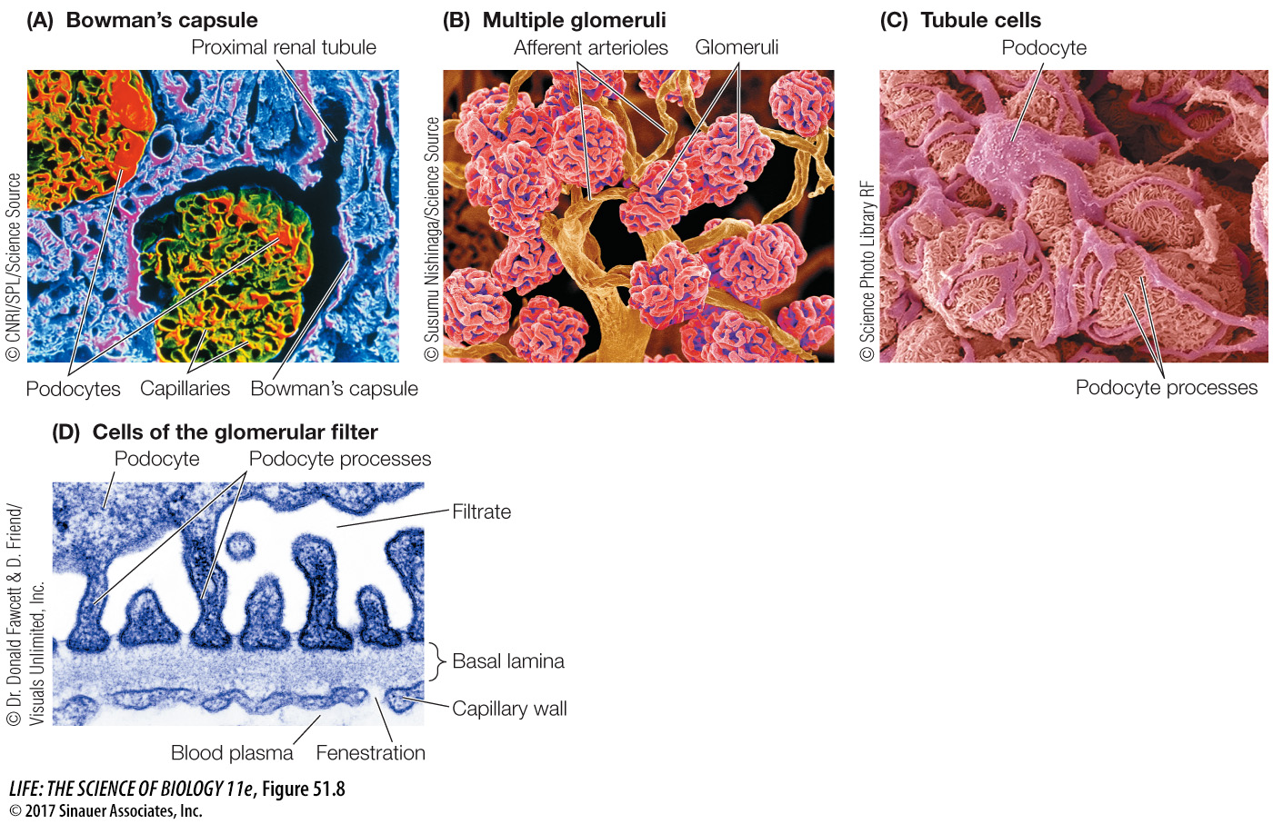
Figure 51.8 A Tour of the Nephron Scanning electron micrographs illustrate the anatomical basis for blood filtration in the kidneys. (A) This cross section of an intact glomerulus shows the tubule cells that form Bowman’s capsule surrounding the glomerular capillaries. (B) In a preparation showing only the blood vessels (tubular tissue has been digested away), the glomeruli appear as balls of capillaries served by arterioles. (C) Higher magnification of a glomerulus with the tubule cells intact shows the podocytes that wrap around the glomerular capillaries. (D) The glomerular filter has three layers: the fenestrated endothelial cells of the capillaries, the meshwork of collagen fibers making up the basement membrane, and the filtration slits between the podocyte processes. All three layers have negative charges, which contribute to their ability to prevent the passage of protein molecules.