Biologists can manipulate living systems to establish cause and effect
How do we know that the structural fibers of the cytoskeleton can achieve all these dynamic functions? We can observe an individual structure under the microscope, and we can observe the functions of living cells that contain that structure. These observations may suggest that the structure carries out a particular function, but mere correlation does not show cause and effect. For example, light microscopy of living cells reveals that the cytoplasm is actively streaming around the cell, and that cytoplasm flows into an extended portion of an amoeboid cell during movement. The observed presence of cytoskeletal components suggests, but does not prove, their role in this process. Science seeks to show the specific links that relate one process, A, to a function, B. In cell biology, two approaches are often used to show that a structure or process A causes function B:
103
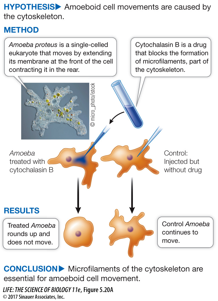
Inhibition: Use a drug that inhibits A and see if B still occurs. If it does not, then A is probably a causative factor for B. Figure 5.20 shows an experiment with such a drug (an inhibitor) that demonstrates cause and effect in the case of the cytoskeleton and cell movement
Mutation: Examine a cell that lacks the gene (or genes) for A and see if B still occurs. If it does not, then A is probably a causative factor for B. Part Four of this book will describe many experiments using this genetic approach.
experiment
Original Paper: Pollard, T. D and R. R. Weihing. 1974. Actin and myosin in cell movement. CRC Critical Reviews of Biochemistry 2: 1–
After a test tube demonstration that the drug cytochalasin B prevented microfilament formation from monomeric precursors, the question was asked: Will the drug work like this in living cells and inhibit cell movement in Amoeba? Complementary experiments showed that the drug did not poison other cellular processes.
work with the data
Figure 5.20B The Role of Microfilaments in Cell Movement—
Original Paper: Pollard, T. D. and R. R. Weihing. 1974.
In a search for natural molecules that have effects on cells—
Several important controls were done to validate the conclusions of the experiment. The experiment was repeated in the presence of the following drugs: cycloheximide, which inhibits new protein synthesis; dinitrophenol, which inhibits new ATP formation (energy); and colchicine, which inhibits the polymerization of microtubules. The results are shown in the table.
| Condition | Rounded cells (%) |
|---|---|
| No drug | 3 |
| Cytochalasin B | 95 |
| Colchicine | 4 |
| Cycloheximide | 3 |
| Cycloheximide + cytochalasin B | 94 |
| Dinitrophenol | 5 |
| Dinitrophenol + cytochalasin B | 85 |
QUESTIONS
Question 1
Explain the reasoning behind each experiment. Why were these controls important?
The reasoning behind these experiments was as follows: if microfilaments are essential for cell movement, movement should not occur in the presence of cytochalasin B; if microtubules are essential for cell movement, movement should not occur in colchicine; if cell movement requires the synthesis of new proteins, movement should not occur in cycloheximide; and if cell movement requires energy, movement should not occur in dinitrophenol. The latter three experiments were important controls to rule out involvement of the three processes involved. The cycloheximide + cytochalasin B and the dinitrophenol + cytochalasin B controls demonstrated that the drugs had independent effects on the cells.
Question 2
Interpret the results of each experiment. What can you conclude about movements in Amoeba and the cytoskeleton?
The experiment with cytochalasin B implicated microfilaments in cell movement. The experiment with colchicine ruled out microtubules. The experiments with cycloheximide showed that new protein synthesis is not necessary for cell movement. The experiments with dinitrophenol showed that cell movement does not require new inputs of energy.
A similar work with the data exercise may be assigned in LaunchPad.