Experiments demonstrate chemiosmosis
Because it is so important, chemiosmosis has been subjected to many lab experiments. Let’s look at two lines of evidence; first, a direct demonstration in the lab that a H+ gradient can drive ATP synthesis, and second, natural disruption of the coupling of electron transport to ATP synthesis.
DIRECT DEMONSTRATION OF CHEMIOSOMOSIS The key experiment that demonstrated that a proton (H+) gradient across a membrane could drive ATP synthesis was first performed using chloroplasts, the organelles in plants that convert the energy in sunlight into chemical energy (photosynthesis; see Chapter 10) (Figure 9.9). Soon thereafter, the same mechanism was shown to work in mitochondria.
experiment
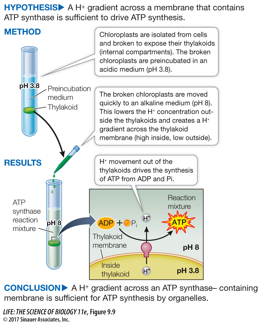
Figure 9.9 An Experiment Demonstrates the Chemiosmotic Mechanism
Original Paper: Jagendorf, A. T. and E. Uribe. 1966. ATP formation caused by acid-
The chemiosmosis hypothesis was a bold departure from the conventional scientific thinking of the time. It required an intact compartment enclosed by a membrane. Could a proton gradient drive the synthesis of ATP? The first experiments to answer this question used chloroplasts, plant organelles that use the same mechanism as mitochondria to synthesize ATP.
A work with the data exercise that accompanies this figure may be assigned in LaunchPad.
Animation 9.2 Two Experiments Demonstrate the Chemiosmotic Mechanism
www.life11e.com/
181
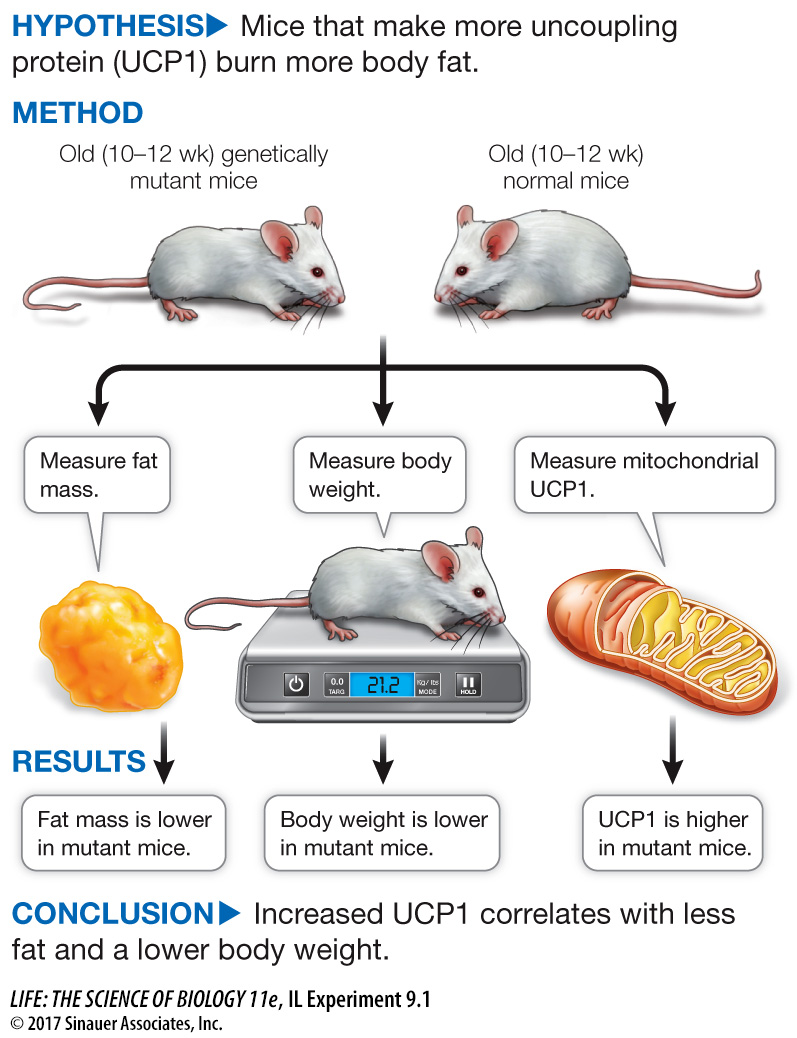
UNCOUPLING ELECTRON TRANSPORT FROM ATP PRODUCTION As you have seen, the coupling of electron transport (which generates the proton gradient) with chemiosmosis is vital for the capture of free energy in the form of ATP. Uncoupling protein 1 (UCP1), which is found in the mitochondria of brown fat cells (described in the chapter opening), demonstrates the importance of this coupling. By disrupting the gradient, UCP1 allows the energy released during electron transport to be in the form of heat, rather than chemical energy trapped in ATP.
The relationship between UCP1 in brown fat and body weight has been investigated experimentally in a study of a genetic strain of mice that—
182
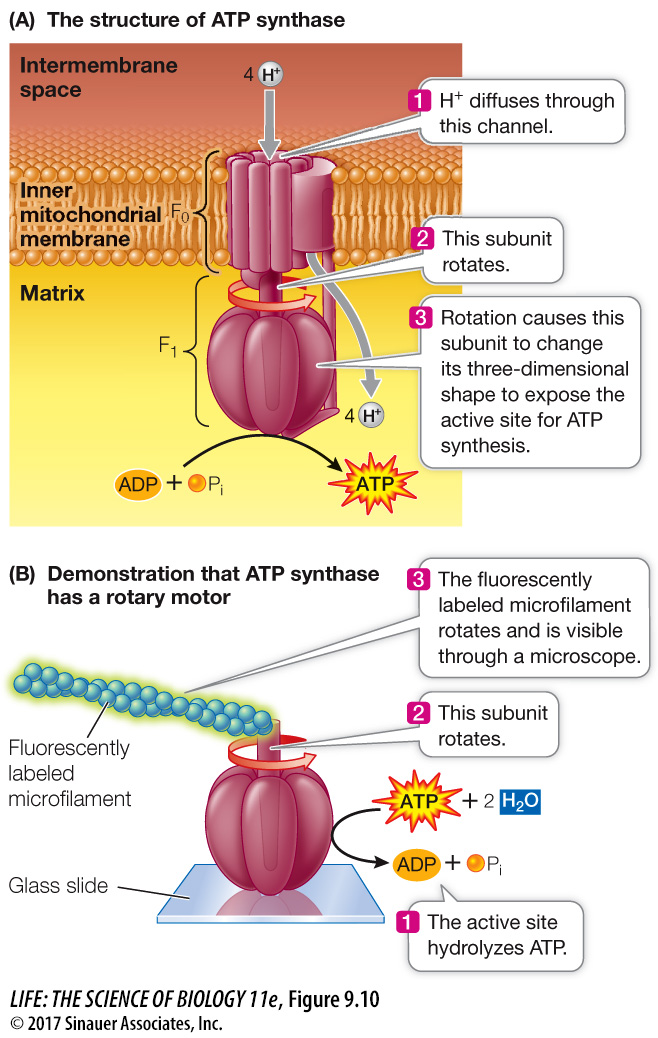
HOW ATP SYNTHASE WORKS: A MOLECULAR MOTOR Now that we have established that the H+ gradient is needed for ATP synthesis, a question remains: How does the enzyme ATP synthase actually make ATP from ADP and Pi? This is certainly a fundamental question in biology, as it underlies energy harvesting in most cells. The structure and mechanism of ATP synthase, illustrated in Figure 9.10A, are shared by living organisms as diverse as bacteria and humans. ATP synthase is a molecular motor composed of two parts: the F0 unit, a transmembrane region that is the H+ channel; and the F1 unit, which contains the active sites for ATP synthesis. F1 consists of six subunits (three each of two polypeptide chains), arranged like the segments of an orange around a central shaftlike polypeptide that interacts with the membrane-
Media Clip 9.1 ATP Synthase in Motion
www.life11e.com/
183
investigating life
Mitochondria, Genetics, and Obesity
experiment
Original Paper: Ma, X., L. Lin, G. Qin, X. Lu, M. Fiorotto, V. Dixit and X. Sun. 2011. Ablations of ghrelin and ghrelin receptor exhibit differential metabolic phenotypes and thermogenic capacity during aging. PLoS One 6: e16391.
As people (and mice) get older, they tend to accumulate fat. In the course of investigating a strain of mice genetically unable to make the receptor for a hormone called ghrelin that is involved in controlling appetite, Yuxiang Sun and her team at Baylor College of Medicine compared these mice with normal mice as they aged. They were surprised to find that the genetically changed mice did not put on as much weight as their normal counterparts. The researchers discovered that the genetic mutation in these mice caused uncoupling of oxidative phosphorylation and burning of body fat. Their experiment investigated whether levels of mitochondrial uncoupling protein 1 (UCP1) were different in the genetically changed mice than in normal mice.
work with the data
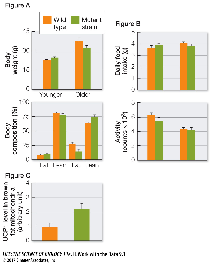
In an effort to determine whether weight gain in normal mice might be due to inactivity or to eating too much (two of the well-
QUESTIONS
Question 1
Did the mutant mice and normal mice gain weight as they aged?
Both groups of mice gained weight as they aged, but the mutant mice gained less weight than the normal mice.
Question 2
The researchers wondered whether the weight changes noted in Figure 1 were due to changes in eating patterns or exercising. So they measured how much food the mice ate per day and measured their movements over time in a special chamber. The results are shown in Figure B. What can you conclude about the role of food intake and exercise on the weight differences between the two strains of mice?
The amount of food eaten and the levels of exercise were the same for both groups. So the lower weight gain in the mutant mice was not due to food intake or exercise.
Question 3
The uncoupling protein UCP1 found in the inner mitochondrial membrane of brown fat cells uncouples mitochondrial electron transport (oxidation) and ATP production (phosphorylation), so that instead of being trapped as chemical energy in the formation of ATP, the energy released by oxidation is released as heat. Sun and her team measured UCP1 levels in brown fat mitochondria of the two strains of mice. The results are shown in Figure C. What can you conclude about the role of UCP1 in the weight differences between the two strains of mice?
UCP1 levels were much higher in the mutant mice than in the normal mice. The correlation between higher UCP1 and lower weight gain indicates that uncoupling of mitochondrial ATP synthesis from electron transport in brown fat may be responsible for lowered weight gain.
A similar work with the data exercise may be assigned in LaunchPad.
184
An ingenious experiment confirmed this rotary motor mechanism. Masasuke Yoshida and his colleagues at the Tokyo Institute of Technology isolated the F1 portion of the ATP synthase and attached it to a glass slide. Fluorescently labeled microfilaments were attached to the central peptide, and the slide was incubated in a solution containing ATP. In this case there was no proton gradient to drive the molecular motor in the direction of ATP synthesis. Instead, ATP was hydrolyzed to ADP and Pi, and this energy caused the motor to spin. Rotation of the labeled microfilament was visible under a microscope, with the labeled filaments clearly rotating like propellers.