key concept 11.3 Eukaryotic Cells Divide by Mitosis
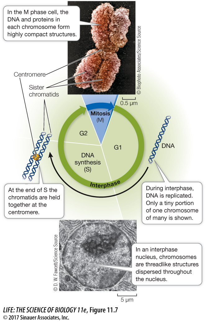
DNA is a very long polymer, up to several µm long. At every stage of the cell cycle, these threads must be packaged into compact structures. A eukaryotic chromosome consists of one or two linear, double-
220
focus your learning
Mitosis ensures that each daughter cell receives one copy of the parent cell’s DNA.
Mitosis involves ordered events within the dividing cell nucleus.
Cytokinesis is the process by which a cell’s cytoplasm divides. It occurs once mitosis is complete.
If all of the DNA in a typical human cell were put end to end, it would be nearly 2 meters long. Yet the nucleus is only 5 µm (0.000005 meters) in diameter and therefore DNA must be extensively packaged in a highly organized way (Figure 11.8). This packing is achieved largely by proteins called histones (histos, “web” or “loom”), which are positively charged at cellular pH because of their high content of the basic amino acids lysine and arginine. The charged R groups on these amino acids bind to the negatively charged phosphate groups on DNA by ionic attractions. These DNA–
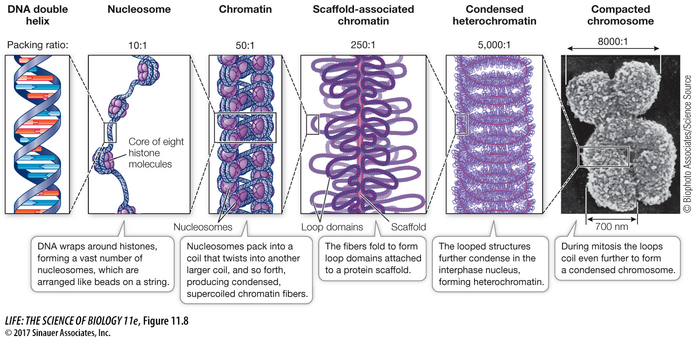
During interphase, the chromatin that makes up each chromosome consists of single DNA molecules running around vast numbers of nucleosomes that resemble beads on a string. During this phase of the cell cycle, the DNA is accessible to proteins involved in replication and transcription. Once a mitotic chromosome is formed, its compact nature makes it inaccessible to replication and transcription factors, and so these processes cannot occur. Further coiling of the chromatin continues up to the time at which the chromatids begin to move apart.
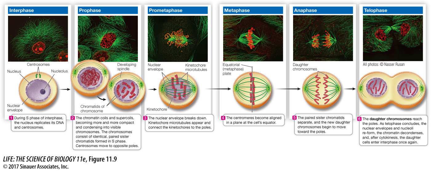
In mitosis, a single nucleus gives rise to two nuclei that are genetically identical to each other and to the parent nucleus. Mitosis (the M phase of the cell cycle) ensures the accurate segregation of the eukaryotic cell’s multiple chromosomes into the daughter nuclei. While mitosis is a continuous process in which each event flows smoothly into the next, it is convenient to subdivide it into a series of stages: prophase, prometaphase, metaphase, anaphase, and telophase (Figure 11.9, Table 11.2).
Activity 11.1 Images of Mitosis
www.Life11e.com/
| Phase | Events |
|---|---|
| Interphase: | |
| G1 | Growth; restriction point at end |
| S | DNA replication |
| G2 | Spindle synthesis begins; preparation for mitosis |
| Mitosis: | |
| Prophase | Condensation of chromosomes; spindle assembly |
| Prometaphase | Nuclear envelope breakdown; chromosome attachment to spindle |
| Metaphase | Alignment of chromosomes at equatorial plate |
| Anaphase | Separation of chromatids; migration to poles |
| Telophase | Chromosomes decondense; nuclear envelope re- |
| Cytokinesis | Cell separation; cell membrane and/or wall formation |
221
Let’s take a closer look at two cellular structures that contribute to the orderly segregation of the chromosomes during mitosis—
Animation 11.1 Mitosis
www.Life11e.com/