Cytokinesis divides the cytoplasm
Cytokinesis divides the cell’s cytoplasm after nuclear division in mitosis. There are substantial differences between the process in animal and plant cells. In animal cells, cytokinesis begins with a furrowing of the cell membrane, as if an invisible thread were cinching the cytoplasm between the two nuclei (Figure 11.12A). This contractile ring is composed of microfilaments of actin and associated myosin (see Figure 5.15), which form a ring on the cytoplasmic surface of the cell membrane. These two proteins interact to produce a contraction, pinching the cell in two. The microfilaments assemble rapidly from actin monomers that are present in the interphase cytoskeleton.
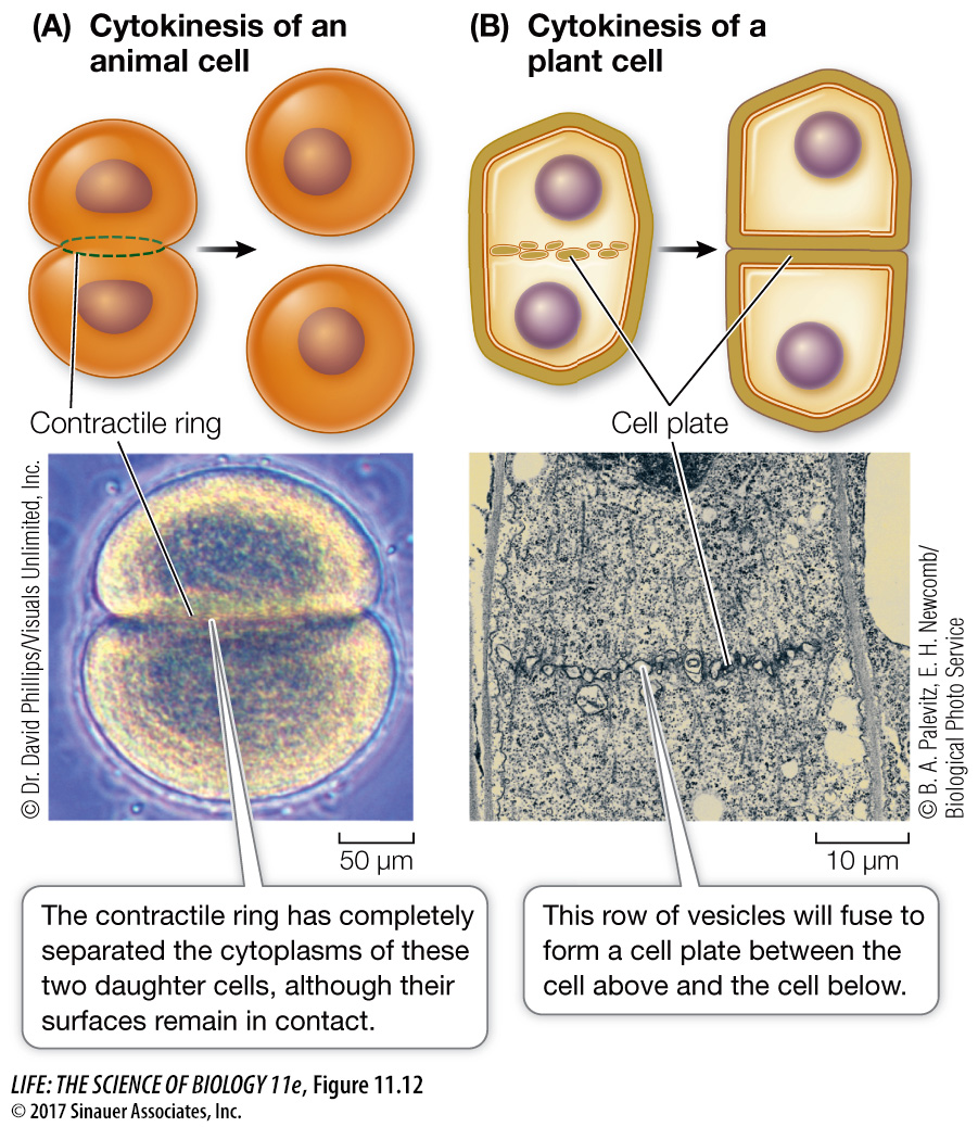
The plant cell cytoplasm divides differently because plants have rigid cell walls. In plant cells, as the spindle breaks down after mitosis, membranous vesicles derived from the Golgi apparatus appear along the plane of cell division, roughly midway between the two daughter nuclei. The vesicles are propelled along microtubules by the motor protein kinesin, and fuse to form a new cell membrane. At the same time they contribute their contents to a cell plate, which is the beginning of a new cell wall (Figure 11.12B).
Following cytokinesis, each daughter cell contains all the components of a complete cell. A precise distribution of chromosomes is ensured by mitosis. In contrast, organelles such as ribosomes, mitochondria, and chloroplasts do not need to be distributed equally between daughter cells, as long as some of each are present in each cell. Accordingly, there is no mechanism with a precision comparable to that of mitosis to provide for their equal allocation to daughter cells.